File:African patient with diabetic retinopathy.jpg

Original file (772 × 976 pixels, file size: 382 KB, MIME type: image/jpeg)
Summary
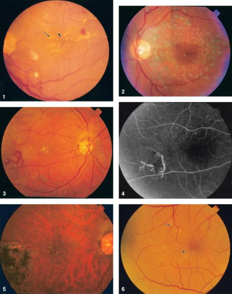
Figure 1. Color fundus photograph of the left eye in case 1 in 1995 showing lipid exudates (arrow), cotton wool spots, and intraretinal hemorrhages of diabetic retinopathy as well as multiple refractile intraretinal crystals (arrowhead). Figure 2. Color fundus photograph of the left eye in case 1 in 2001 after focal photocoagulation for diabetic macular edema. The edema has completely resolved, and there are fewer intraretinal crystals, which are arranged in a pattern different from that in 1995. Figure 3. Color fundus photograph of the right eye in case 2 showing a frond of retinal neovascularization and many iridescent intraretinal crystals within the foveal avascular zone. Figure 4. Mid-phase fluorescein angiogram of the right eye in case 2 showing the perfused retinal neovascularization and the absence of any fluorescein angiographic sign of the crystals. Figure 5. Color fundus photograph of the right eye in case 2 one year after panretinal laser photocoagulation. Most of the crystals have resolved. Figure 6. Color fundus photograph of the right eye in case 3. The distribution of the intraretinal crystals follows the pattern of the distribution of intraretinal lipid from diabetic macular edema. The diabetic lipid exudates (arrow) can be distinguished from the refractile crystals (arrowhead). West African Crystalline Maculopathy
Ophthalmology 2004;111:921–925 © 2004
File history
Click on a date/time to view the file as it appeared at that time.
| Date/Time | Thumbnail | Dimensions | User | Comment | |
|---|---|---|---|---|---|
| current | 13:11, May 5, 2023 | | 772 × 976 (382 KB) | Tony.Ching.AAO (talk | contribs) | Figure 1. Color fundus photograph of the left eye in case 1 in 1995 showing lipid exudates (arrow), cotton wool spots, and intraretinal hemorrhages of diabetic retinopathy as well as multiple refractile intraretinal crystals (arrowhead). Figure 2. Color fundus photograph of the left eye in case 1 in 2001 after focal photocoagulation for diabetic macular edema. The edema has completely resolved, and there are fewer intraretinal crystals, which are arranged in a pattern different from that in 1... |
You cannot overwrite this file.
File usage
The following page uses this file: