

File:CNV Type 2.jpg
From EyeWiki
No higher resolution available.
CNV_Type_2.jpg (670 × 540 pixels, file size: 122 KB, MIME type: image/jpeg)
Summary
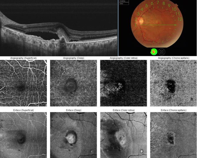
Type 2 CNV. In B-scan and OCT-A, a neovascular lesion is identified that extends from the choroidal vessels through the Bruch's membrane and RPE and grows into the subretinal space. The En Face image shows a change in color in macula secondary to edema and serous detachment of the retina. (Courtesy of Andrea Tamine Hoyos Dumar, MD.)
File history
Click on a date/time to view the file as it appeared at that time.
| Date/Time | Thumbnail | Dimensions | User | Comment | |
|---|---|---|---|---|---|
| current | 00:48, June 3, 2017 | | 670 × 540 (122 KB) | Dan.Mummert.AAO (talk | contribs) |
You cannot overwrite this file.
File usage
The following page uses this file:


