

File:Conjunctival amyloidosis gross photo.png
From EyeWiki
No higher resolution available.
Conjunctival_amyloidosis_gross_photo.png (512 × 305 pixels, file size: 284 KB, MIME type: image/png)
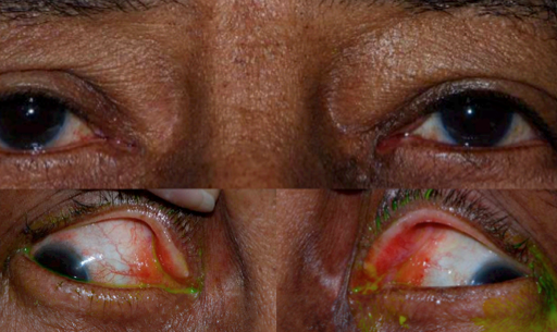
Figure 2: Conjunctival amyloidosis. Smooth, waxy, yellow, relatively circumscribed masses are seen at bilateral medial canthi. Visualization of the lesions is improved with lateral gaze. (Image courtesy of Gary S. Lissner, MD)
File history
Click on a date/time to view the file as it appeared at that time.
| Date/Time | Thumbnail | Dimensions | User | Comment | |
|---|---|---|---|---|---|
| current | 15:15, January 26, 2014 | | 512 × 305 (284 KB) | Frank.Hrisomalos (talk | contribs) | Figure 2: Conjunctival amyloidosis. Smooth, waxy, yellow, relatively circumscribed masses are seen at bilateral medial canthi. Visualization of the lesions is improved with lateral gaze. (Image courtesy of Gary S. Lissner, MD) |
You cannot overwrite this file.
File usage
The following page uses this file:


