File:FAF.gyrateatrophy.PNG
From EyeWiki
Size of this preview: 800 × 577 pixels. Other resolution: 1,995 × 1,439 pixels.
Original file (1,995 × 1,439 pixels, file size: 1.52 MB, MIME type: image/png)
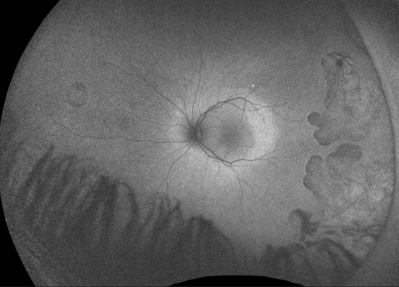
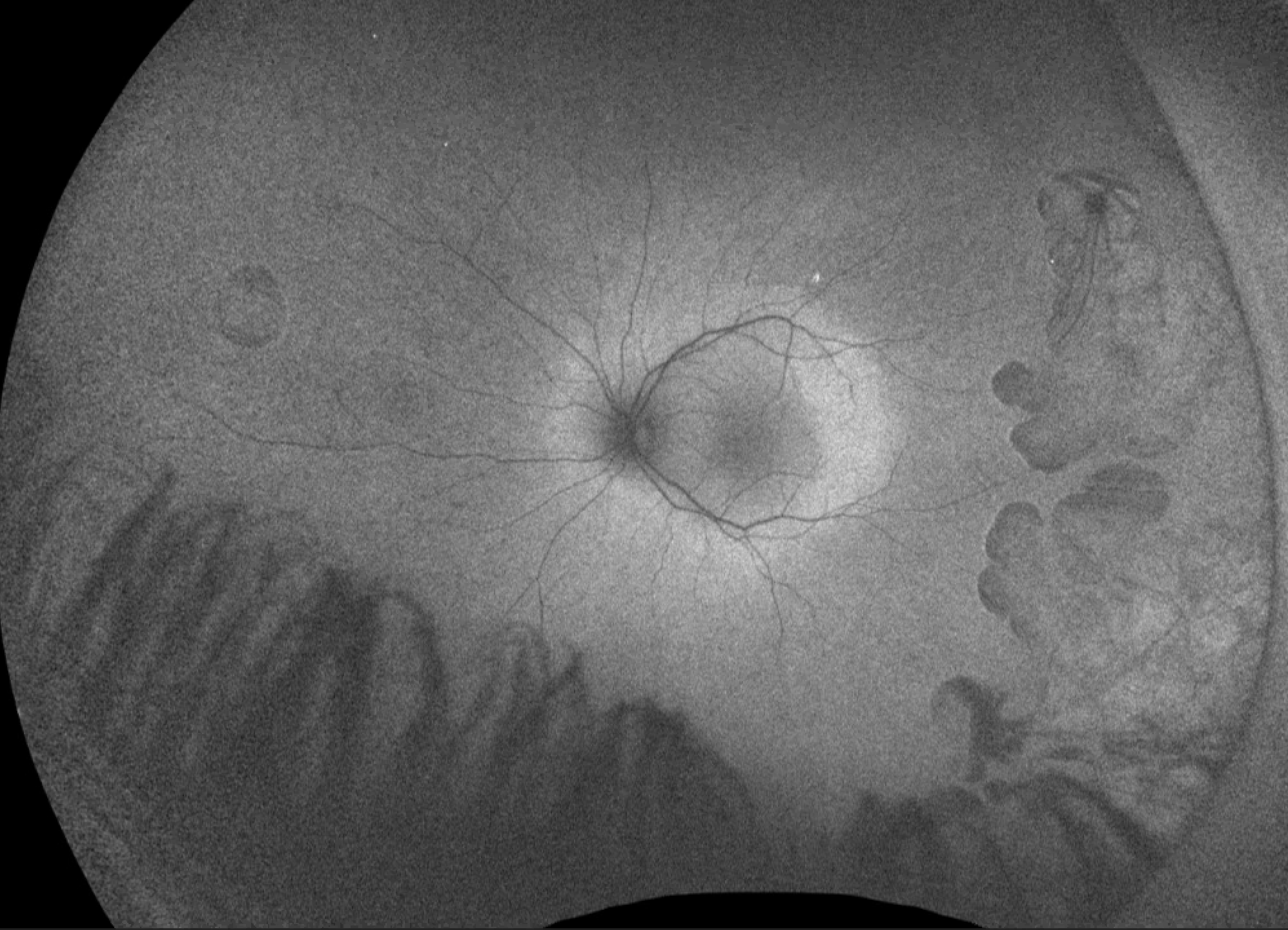
Fundus autofluorescence of a patient with early gyrate atrophy showing peripheral hypoautofluorescent lesions correlating with chorioretinal atrophy seen on exam. Note the thin demarcated hyperautofluorescent perimeter to such lesions. Photo courtesy of Dr. Marc Mathias.
File history
Click on a date/time to view the file as it appeared at that time.
| Date/Time | Thumbnail | Dimensions | User | Comment | |
|---|---|---|---|---|---|
| current | 10:10, April 9, 2019 | | 1,995 × 1,439 (1.52 MB) | Eric.Weldy (talk | contribs) | Fundus autofluorescence of a patient with early gyrate atrophy showing peripheral absence of autofluorescence correlating with chorioretinal atrophy seen on exam. Note the thin demarcated hyperautofluorescent perimeter to such lesions. The macula sh... |
You cannot overwrite this file.
File usage
The following page uses this file: