File:Figure 1A.jpg
From EyeWiki
Size of this preview: 700 × 599 pixels. Other resolution: 2,392 × 2,048 pixels.
Original file (2,392 × 2,048 pixels, file size: 825 KB, MIME type: image/jpeg)
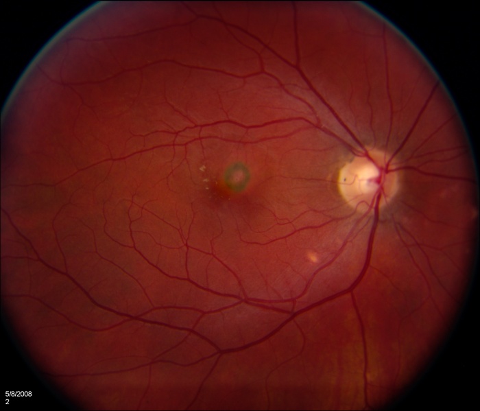
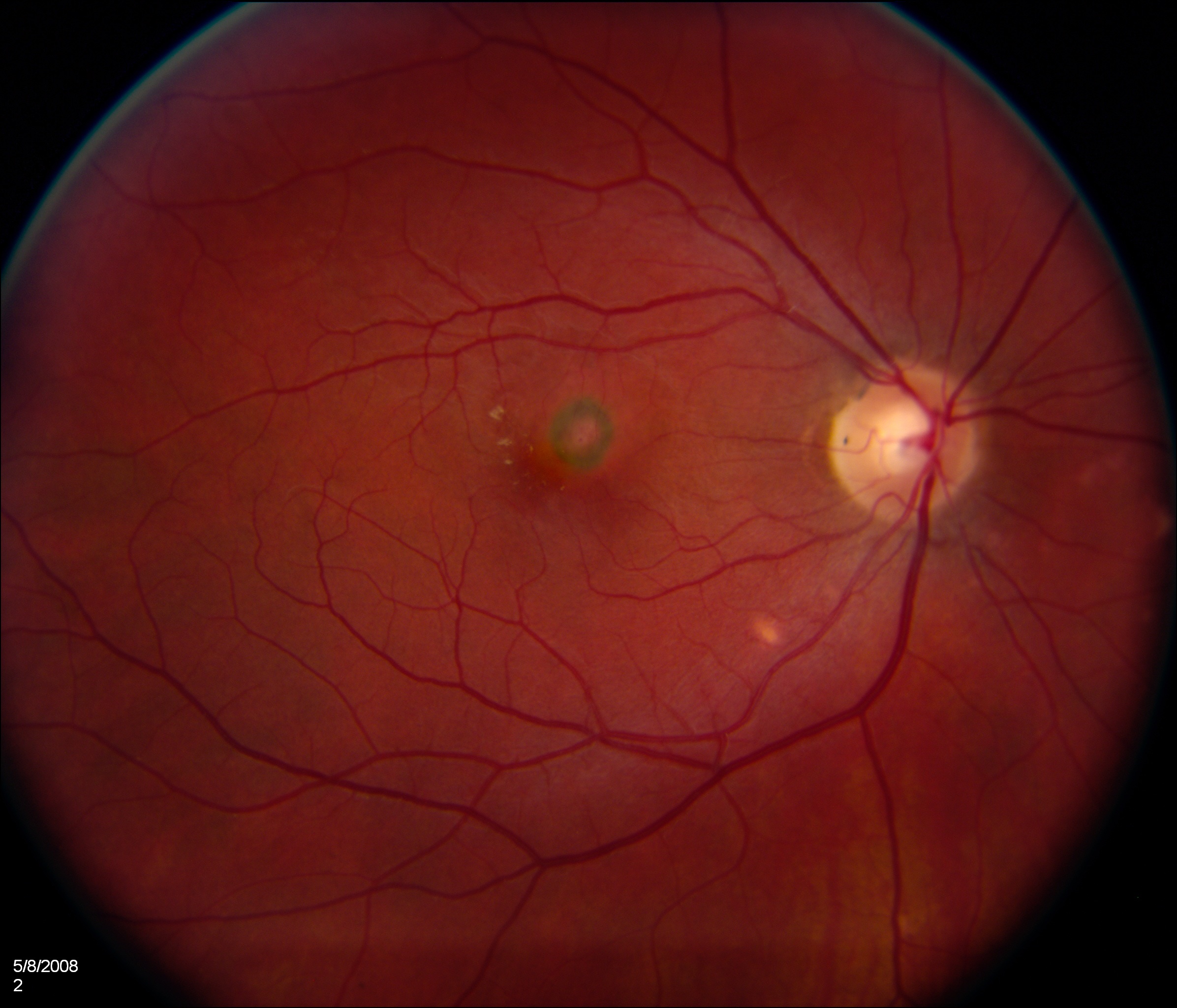
Preoperative color photograph demonstrates multiple, small, yellowish punctate lesions at the level of the choroid. A subfoveal CNV membrane with hyperpigmented borders is seen.
File history
Click on a date/time to view the file as it appeared at that time.
| Date/Time | Thumbnail | Dimensions | User | Comment | |
|---|---|---|---|---|---|
| current | 18:42, November 30, 2011 | | 2,392 × 2,048 (825 KB) | Katrina.A.Mears (talk | contribs) | Preoperative color photograph demonstrates multiple, small, yellowish punctate lesions at the level of the choroid. A subfoveal CNV membrane with hyperpigmented borders is seen. Courtesy of Dr Narsing Rao and taken from the following publication: Pachyd |
| 18:38, November 30, 2011 | 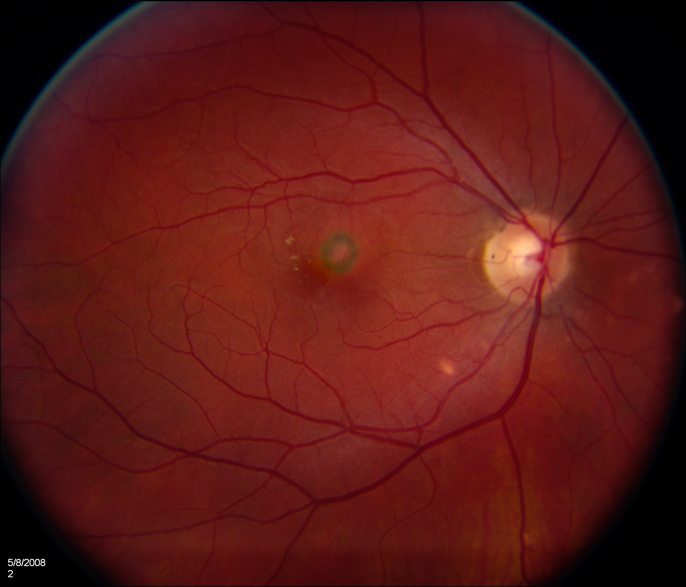 | 2,392 × 2,048 (825 KB) | Katrina.A.Mears (talk | contribs) | Preoperative color photograph demonstrates multiple, small, yellowish punctate lesions at the level of the choroid. A subfoveal CNV membrane with hyperpigmented borders is seen. |
You cannot overwrite this file.
File usage
The following page uses this file: