File:Figure 1 MRI.jpg
From EyeWiki
Size of this preview: 800 × 401 pixels. Other resolution: 1,096 × 550 pixels.
Original file (1,096 × 550 pixels, file size: 180 KB, MIME type: image/jpeg)
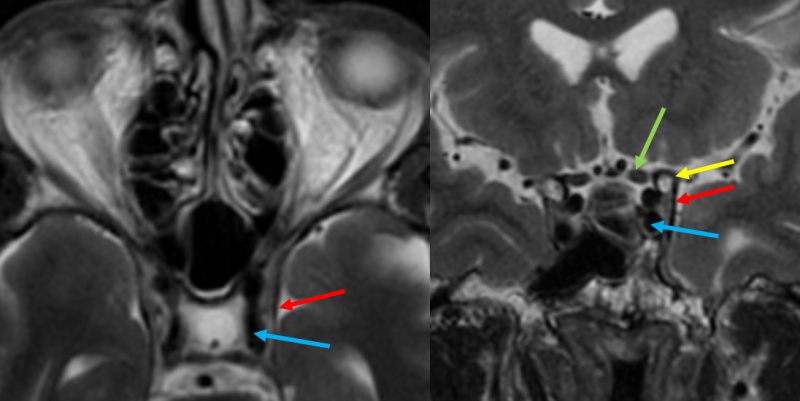
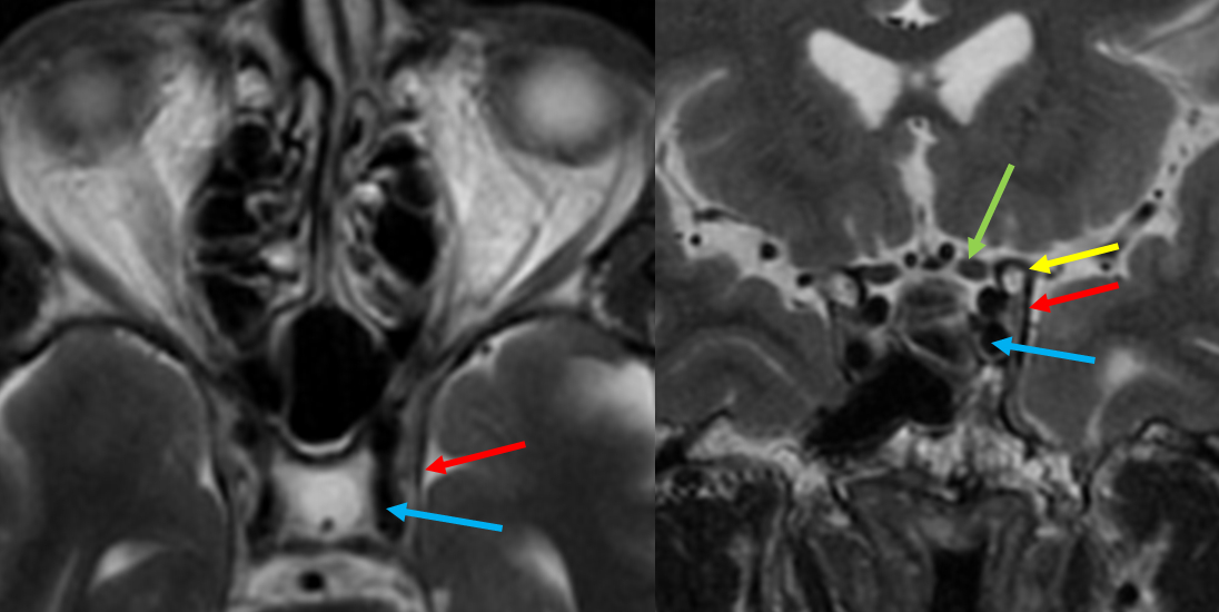
Figure 1: MRI T2 sequence, axial (left) and coronal (right) views, shows the horizontal segment of the left cavernous ICA (blue arrow), lateral dural wall (red), anterior clinoid process (ACP, yellow) and optic nerve (green). Coronal view also shows the clinoid segment of the ICA in cross-section. Image courtesy of John D. Nerva, MD.
File history
Click on a date/time to view the file as it appeared at that time.
| Date/Time | Thumbnail | Dimensions | User | Comment | |
|---|---|---|---|---|---|
| current | 14:04, March 27, 2020 | | 1,096 × 550 (180 KB) | Jacquelyn.Laplant (talk | contribs) | Figure 1: MRI T2 sequence, axial (left) and coronal (right) views, shows the horizontal segment of the left cavernous ICA (blue arrow), lateral dural wall (red), anterior clinoid process (ACP, yellow) and optic nerve (green). Coronal view also shows th... |
You cannot overwrite this file.
File usage
The following page uses this file: