File:Figure 2.jpg
From EyeWiki
Size of this preview: 800 × 428 pixels. Other resolution: 3,557 × 1,902 pixels.
Original file (3,557 × 1,902 pixels, file size: 427 KB, MIME type: image/jpeg)
File history
Click on a date/time to view the file as it appeared at that time.
| Date/Time | Thumbnail | Dimensions | User | Comment | |
|---|---|---|---|---|---|
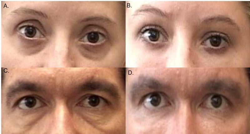
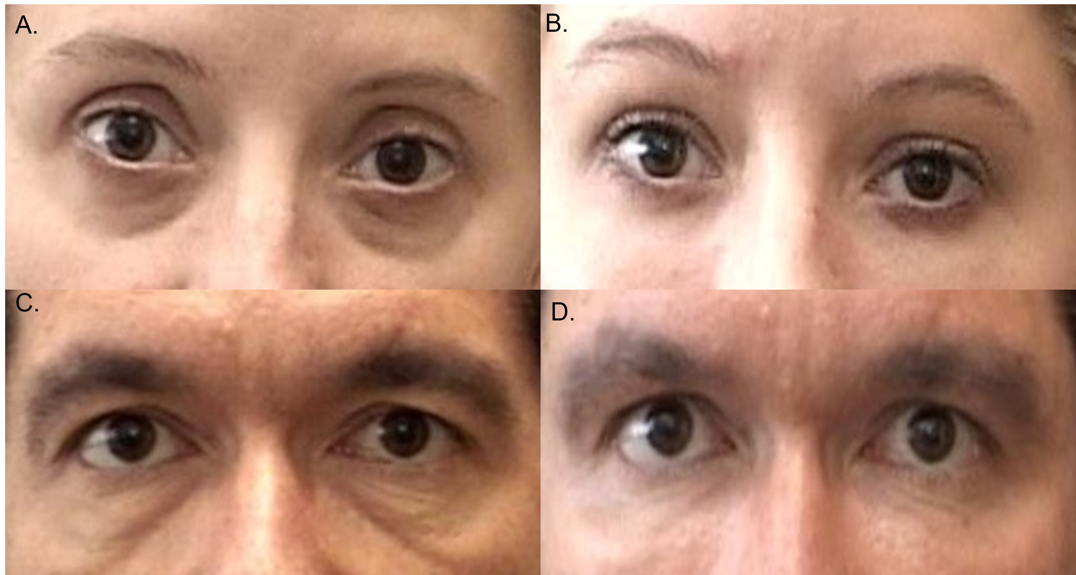
| current | 16:14, June 20, 2023 | | 3,557 × 1,902 (427 KB) | Jan.Ulloa (talk | contribs) | Figure 2: Tear through deformity treated with fat grafting. A, Pre-fat grafting of patient 1. B, Post-fat grafting of patient 1. C, Pre-fat grafting of patient 2. D, Post-fat grafting of patient 2. |
| 11:45, April 30, 2020 | 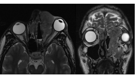 | 438 × 242 (65 KB) | Sarah.Avila (talk | contribs) | MRI showing a large left-sided nasal cavity mass with intracranial, retropharyngeal and orbital extension. There is a notable mass effect on the globe causing compression, proptosis and stretching of the optic nerve. Biopsy confirmed esthesioneuroblas... | |
| 07:03, April 1, 2013 |  | 2,721 × 5,020 (645 KB) | Seyed-Farzad (talk | contribs) | ||
| 23:05, November 25, 2011 |  | 450 × 338 (82 KB) | Lespandar (talk | contribs) |
You cannot overwrite this file.
File usage
The following file is a duplicate of this file (more details):
There are no pages that use this file.