File:Figure 2 Circinate Exudates.jpg
From EyeWiki
Size of this preview: 473 × 600 pixels. Other resolution: 867 × 1,099 pixels.
Original file (867 × 1,099 pixels, file size: 172 KB, MIME type: image/jpeg)
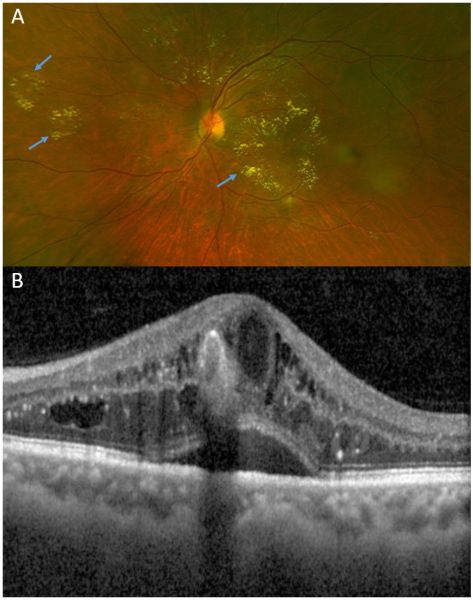
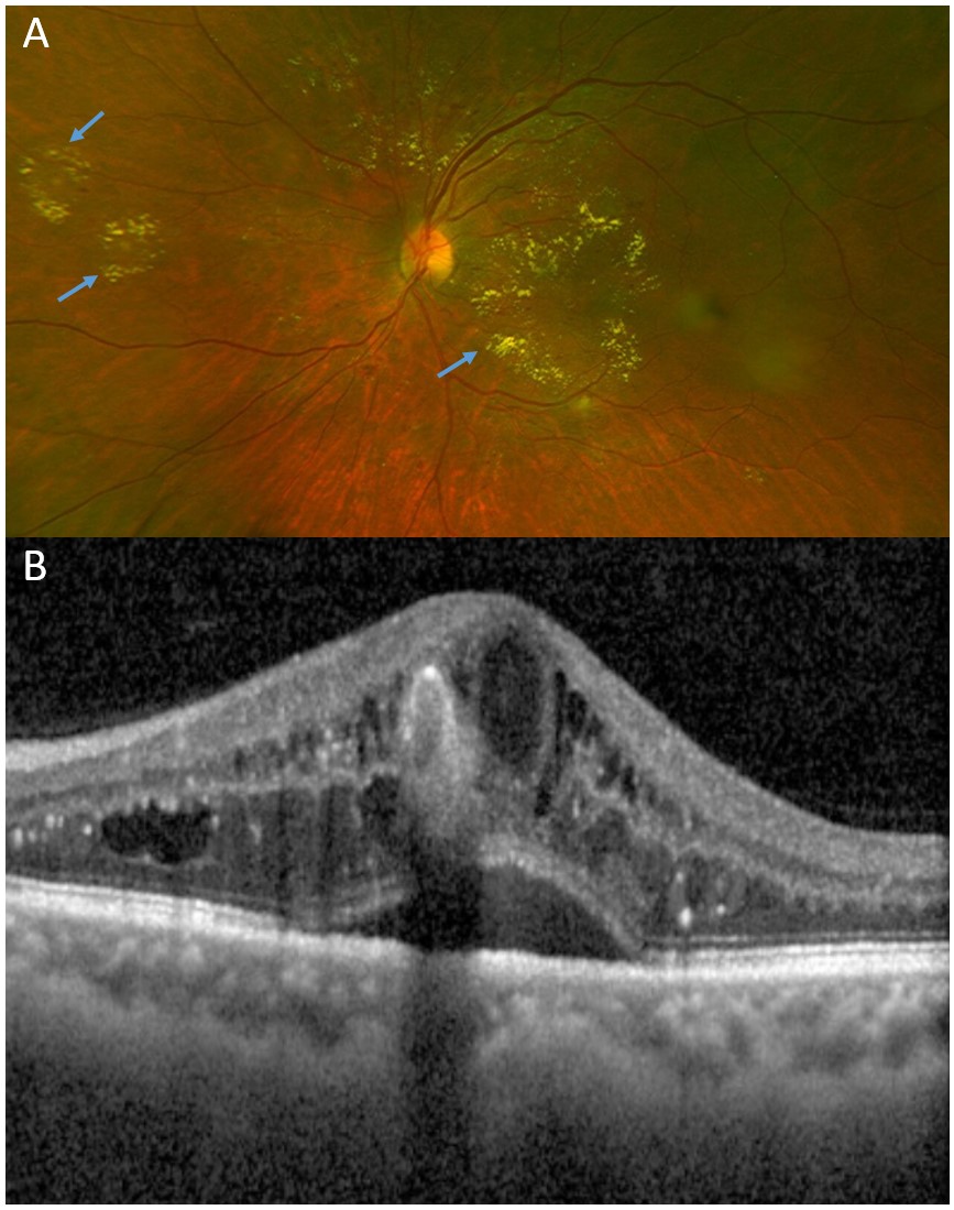
A: Blue arrows - Circinate exudates, defined as exudates arranged in a circular appearance. B: OCT b-scan across the fovea confirming the presence of DME associated with an elevated foveal contour. The patient’s best-corrected vision was 20/32.
File history
Click on a date/time to view the file as it appeared at that time.
| Date/Time | Thumbnail | Dimensions | User | Comment | |
|---|---|---|---|---|---|
| current | 12:46, February 23, 2022 | | 867 × 1,099 (172 KB) | Cris.Jacoba (talk | contribs) |
You cannot overwrite this file.
File usage
The following page uses this file: