File:Figure 3A.jpg
From EyeWiki
Size of this preview: 700 × 599 pixels. Other resolution: 2,392 × 2,048 pixels.
Original file (2,392 × 2,048 pixels, file size: 1.45 MB, MIME type: image/jpeg)
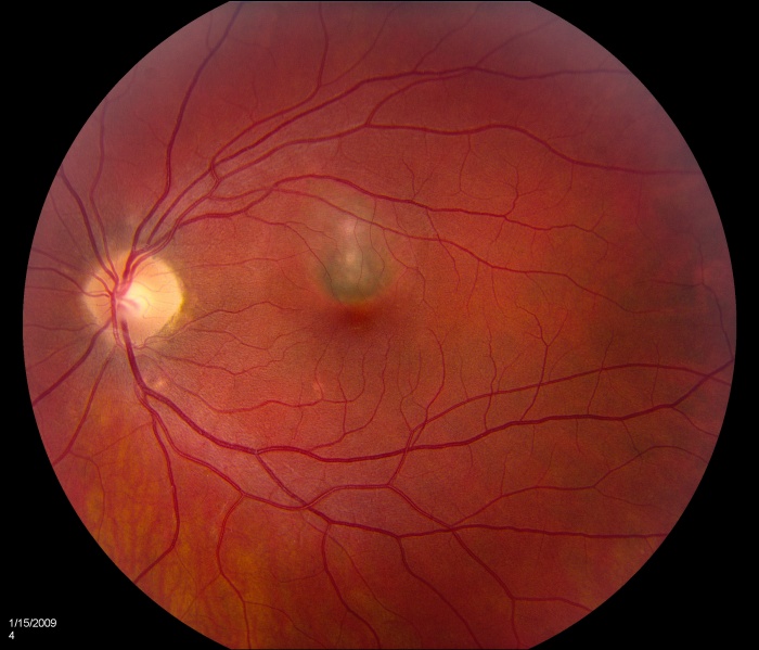
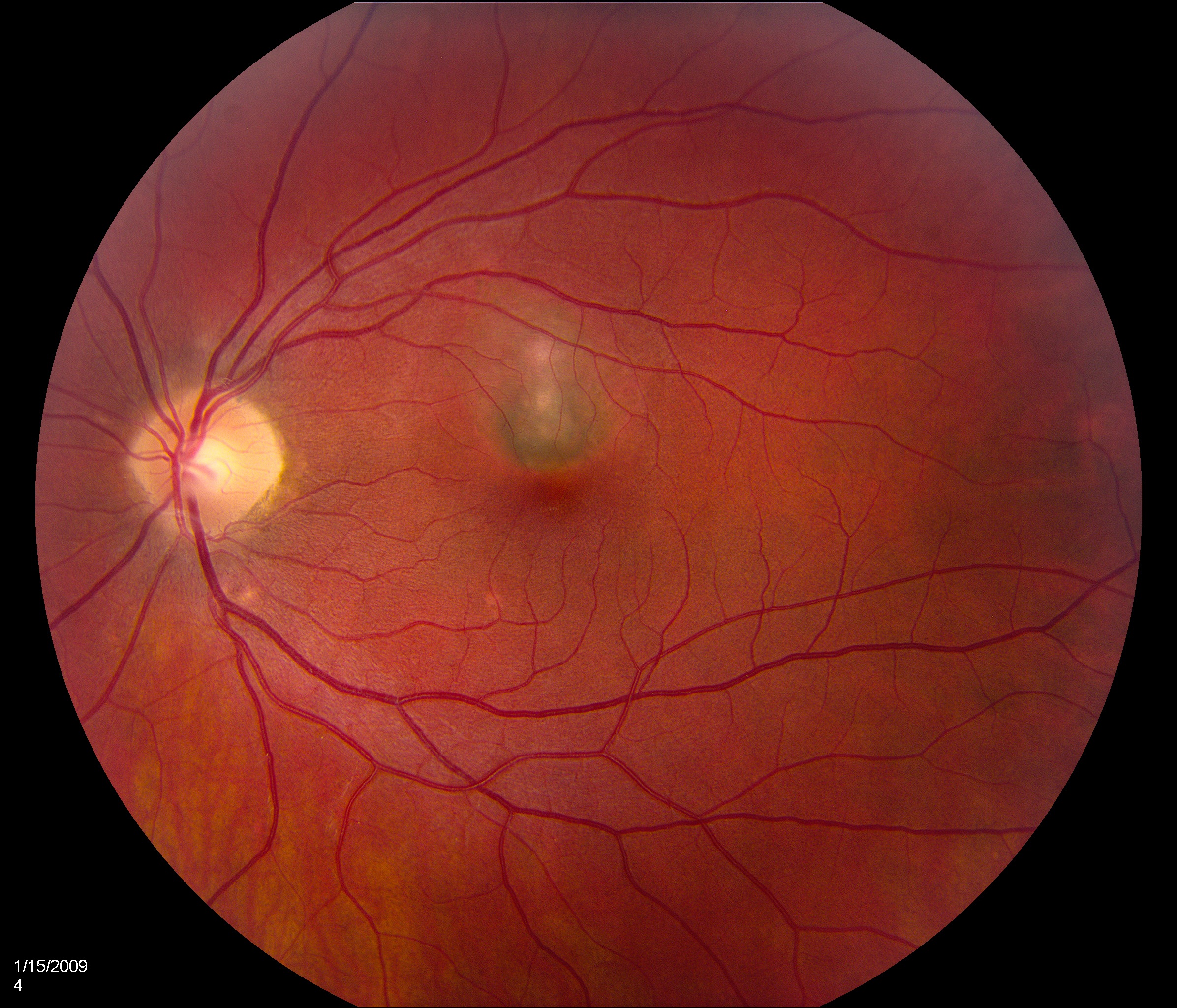
Color fundus photograph 3 months after second injection of bevacizumab shows a grayish subfoveal CNV membrane.
Courtesy of Dr Narsing Rao and taken from the following publication:
Pachydaki SI, Jakobiec FA, Bhat P, Sobrin L, Michaud NA, Seshan SV, D'Amico DJ. Surgical management and ultrastructural study of choroidal neovascularization in punctate inner choroidopathy after bevacizumab. J Ophthalmic Inflamm Infect. 2011 Nov 27.
File history
Click on a date/time to view the file as it appeared at that time.
| Date/Time | Thumbnail | Dimensions | User | Comment | |
|---|---|---|---|---|---|
| current | 21:14, November 30, 2011 | | 2,392 × 2,048 (1.45 MB) | Katrina.A.Mears (talk | contribs) | Color fundus photograph 3 months after second injection of bevacizumab shows a grayish subfoveal CNV membrane. Courtesy of Dr Narsing Rao and taken from the following publication: Pachydaki SI, Jakobiec FA, Bhat P, Sobrin L, Michaud NA, Seshan SV, D'Am |
You cannot overwrite this file.
File usage
The following page uses this file: