

File:Figure 4 - Diabetic macular ischemia by fluorescein angiography.jpg
From EyeWiki
Size of this preview: 743 × 600 pixels. Other resolution: 1,556 × 1,256 pixels.
Original file (1,556 × 1,256 pixels, file size: 411 KB, MIME type: image/jpeg)
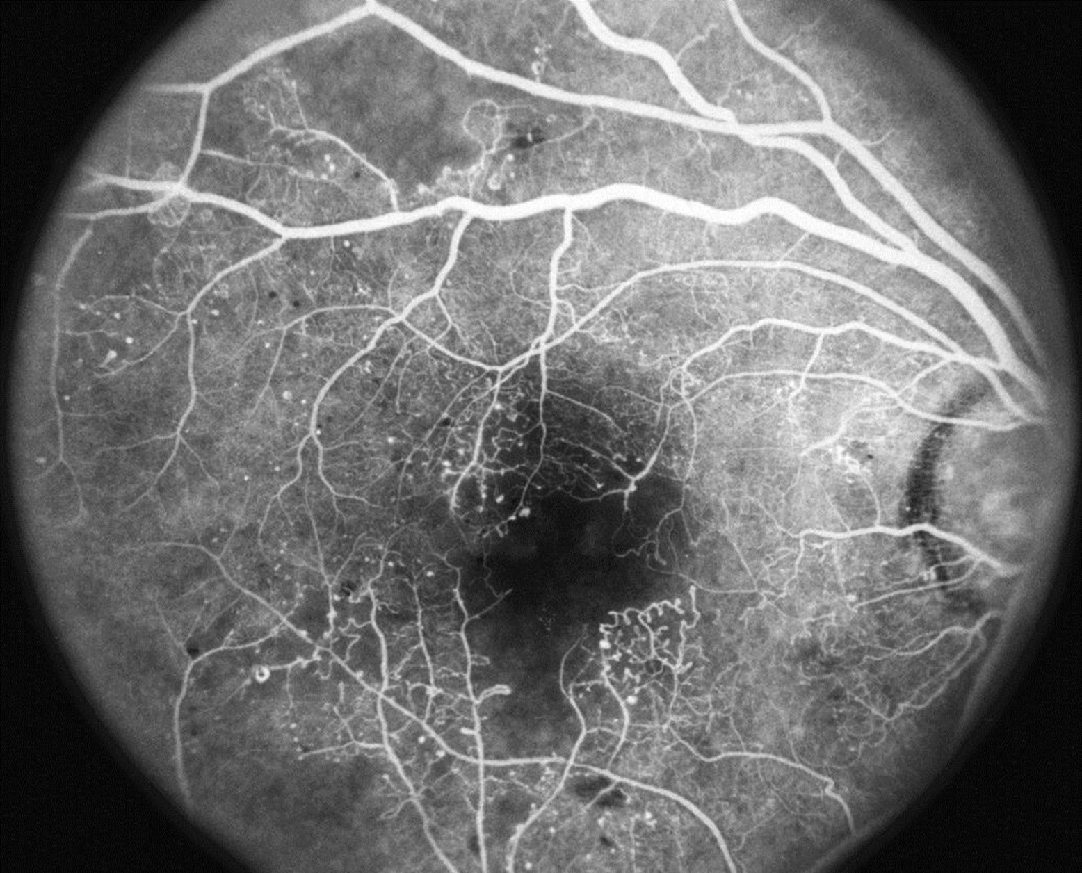
Fluorescein angiography of a diabetic patient with decreased vision due to poor perfusion of the capillary net at the macula. Image shown is in the arterio-venous phase. Large area of macular nonperfusion of about 1-disc diameter is evident. This is seen as a large dark area at the center of macula. Capillary dilatation surrounding the enlarged FAZ is also appreciated, along with large areas of non-perfusion along the superior and inferior arcade. Published online with permission from the AAO.
File history
Click on a date/time to view the file as it appeared at that time.
| Date/Time | Thumbnail | Dimensions | User | Comment | |
|---|---|---|---|---|---|
| current | 13:22, October 20, 2021 | | 1,556 × 1,256 (411 KB) | Cris.Jacoba (talk | contribs) |
You cannot overwrite this file.
File usage
The following page uses this file:


