

File:Figure 5 ETDRS standard photos for diabetic macular ischemia.jpg
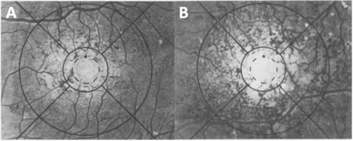
Figure_5_ETDRS_standard_photos_for_diabetic_macular_ischemia.jpg (700 × 281 pixels, file size: 120 KB, MIME type: image/jpeg)
Figure 5: ETDRS standard photos for diabetic macular ischemia. A: Standard photograph 1A (left eye). The central and inner nasal subfield provides the lower boundary for grade 3 (moderate, M) capillary loss. B: Standard photograph 2 (left eye). The central and inner temporal subfields provide the lower boundary for grade 4 (severe, S) capillary loss. In the central subfield, the few capillaries that remain represent 10 to 25% of the number that might be expected. Reprinted from: Classification of diabetic retinopathy from fluorescein angiograms: ETDRS report number 11. Ophthalmology. 1991;98(5):807-822. Published online with permission from Ophthalmology/Copyright Clearance Center.
File history
Click on a date/time to view the file as it appeared at that time.
| Date/Time | Thumbnail | Dimensions | User | Comment | |
|---|---|---|---|---|---|
| current | 09:16, October 27, 2021 | | 700 × 281 (120 KB) | Cris.Jacoba (talk | contribs) |
You cannot overwrite this file.
File usage
The following page uses this file:


