

File:Figure 6 Disorganization of the Retinal Inner Layers.jpg
From EyeWiki
Size of this preview: 800 × 502 pixels. Other resolution: 1,105 × 693 pixels.
Original file (1,105 × 693 pixels, file size: 160 KB, MIME type: image/jpeg)
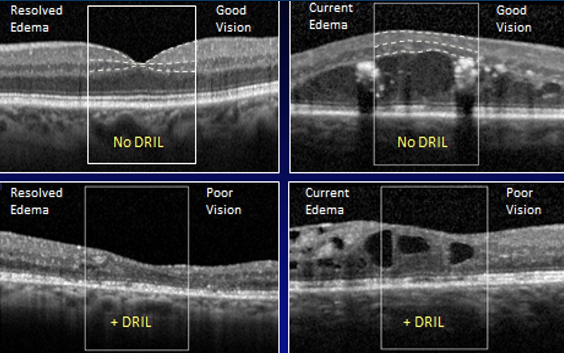
OCT images showing combinations of presence or absence of DRIL and DME. The central 1-mm area is enclosed in the box. Upper images show segmentation of inner retinal layers, with the white lines demarcating the boundaries between IPL-INL and the INL-OPL. Image credit to: Barham et al., Association of Macular Nonperfusion, Leakage, and DRIL (ARVO poster, 2016)
File history
Click on a date/time to view the file as it appeared at that time.
| Date/Time | Thumbnail | Dimensions | User | Comment | |
|---|---|---|---|---|---|
| current | 12:59, February 23, 2022 | | 1,105 × 693 (160 KB) | Cris.Jacoba (talk | contribs) |
You cannot overwrite this file.
File usage
The following page uses this file:


