

File:Figure 9 AS OCT.jpeg
From EyeWiki
No higher resolution available.
Figure_9_AS_OCT.jpeg (624 × 175 pixels, file size: 20 KB, MIME type: image/jpeg)
Summary
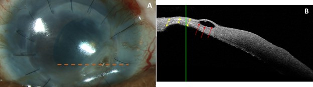
Figure 9A: Slit lamp photo of the right eye showing stromal thinning, scarring and penetrating keratoplasty graft with intact sutures. The dashed orange line in 9A represents the cross-section of the AS-OCT image in 9B. Figure 9B: AS-OCT highlighting the corneal thinning without perforation (yellow double arrows) along with a sub-epithelial pseudocyst (red arrows).
File history
Click on a date/time to view the file as it appeared at that time.
| Date/Time | Thumbnail | Dimensions | User | Comment | |
|---|---|---|---|---|---|
| current | 18:19, October 18, 2022 | | 624 × 175 (20 KB) | Justin.Yamanuha (talk | contribs) | Figure 9A: Slit lamp photo of the right eye showing stromal thinning, scarring and penetrating keratoplasty graft with intact sutures. The dashed orange line in 9A represents the cross-section of the AS-OCT image in 9B. Figure 9B: AS-OCT highlighting the corneal thinning without perforation (yellow double arrows) along with a sub-epithelial pseudocyst (red arrows). |
You cannot overwrite this file.
File usage
The following page uses this file:


