

File:Funduscopic Image Figure 3.png
From EyeWiki
No higher resolution available.
Funduscopic_Image_Figure_3.png (495 × 485 pixels, file size: 346 KB, MIME type: image/png)
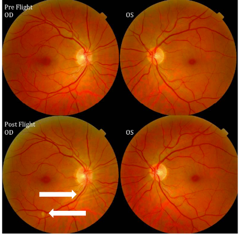
Figure 3: Pre-flight and Post-flight fundoscopic images from LDAF. Post-flight examination reveals choroidal folds (right-pointing arrow) and a single cotton-wool spot (left-pointing arrow). Source: NASA Risk of Spaceflight Associated Neuro-ocular Syndrome Evidence Report
File history
Click on a date/time to view the file as it appeared at that time.
| Date/Time | Thumbnail | Dimensions | User | Comment | |
|---|---|---|---|---|---|
| current | 16:47, December 13, 2020 | | 495 × 485 (346 KB) | Peter.Mortensen (talk | contribs) | Figure 3: Pre-flight and Post-flight fundoscopic images from LDAF. Post-flight examination reveals choroidal folds (right-pointing arrow) and a single cotton-wool spot (left-pointing arrow). Source: NASA Risk of Spaceflight Associated Neuro-ocular Syn... |
You cannot overwrite this file.
File usage
The following page uses this file:


