File:Gyrateatrophy3.PNG
From EyeWiki
Size of this preview: 800 × 552 pixels. Other resolution: 2,052 × 1,416 pixels.
Original file (2,052 × 1,416 pixels, file size: 1.61 MB, MIME type: image/png)
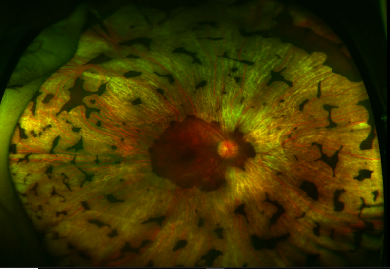
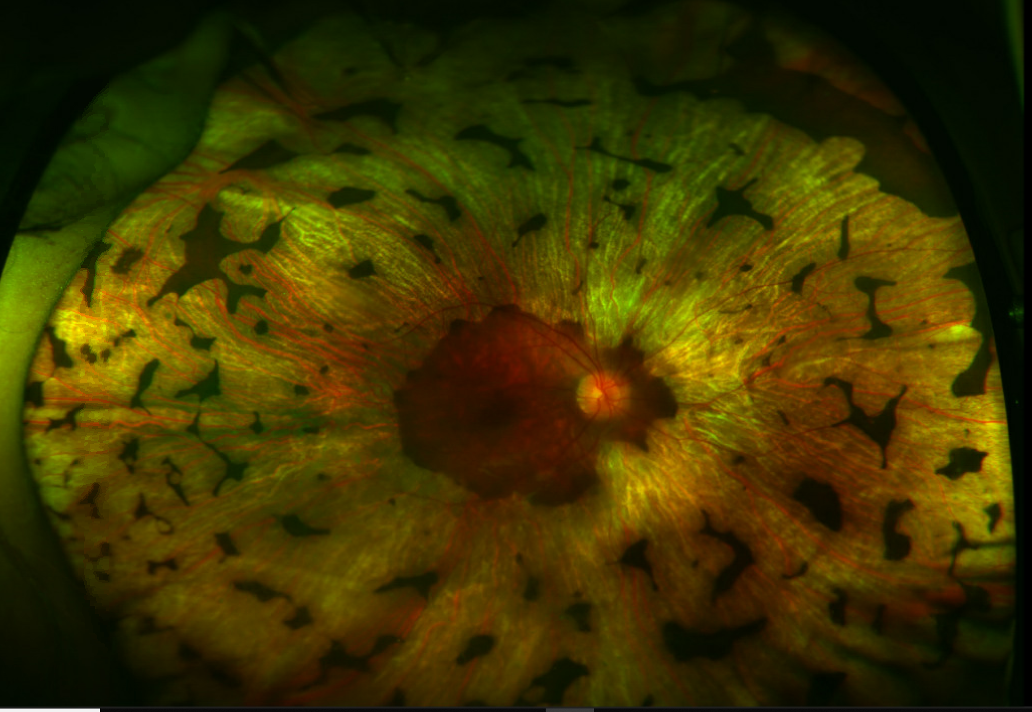
Wide field fundus photography showing an advanced case of gyrate atrophy. Previously circular atrophic lesions have coalesced to form a large confluent area of atrophy with late preservation for macula. The hyperpigmented lesions scattered throughout the periphery appear to mimic bony spicules as seen in retinitis pigmentosa but actually represents islands of relatively unaffected residual retinal tissue that have not yet become confluent. Note the hyperpigmentation of the macula. Photo courtesy of Dr. Marc Mathias, MD.
File history
Click on a date/time to view the file as it appeared at that time.
| Date/Time | Thumbnail | Dimensions | User | Comment | |
|---|---|---|---|---|---|
| current | 09:58, April 9, 2019 | | 2,052 × 1,416 (1.61 MB) | Eric.Weldy (talk | contribs) | Wide field fundus photography showing an advanced case of gyrate atrophy. Previously circular atrophic lesions have coalesced to form a large confluent area of atrophy with late preservation for macula. The hyperpigmented lesions scattered throughout... |
You cannot overwrite this file.
File usage
The following page uses this file: