File:Lymphangioma.png
From EyeWiki
Size of this preview: 800 × 501 pixels. Other resolution: 2,191 × 1,372 pixels.
Original file (2,191 × 1,372 pixels, file size: 2.45 MB, MIME type: image/png)
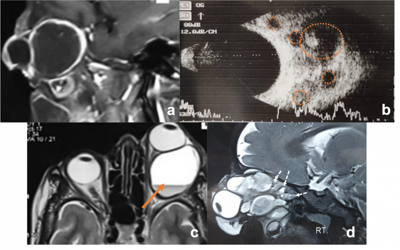
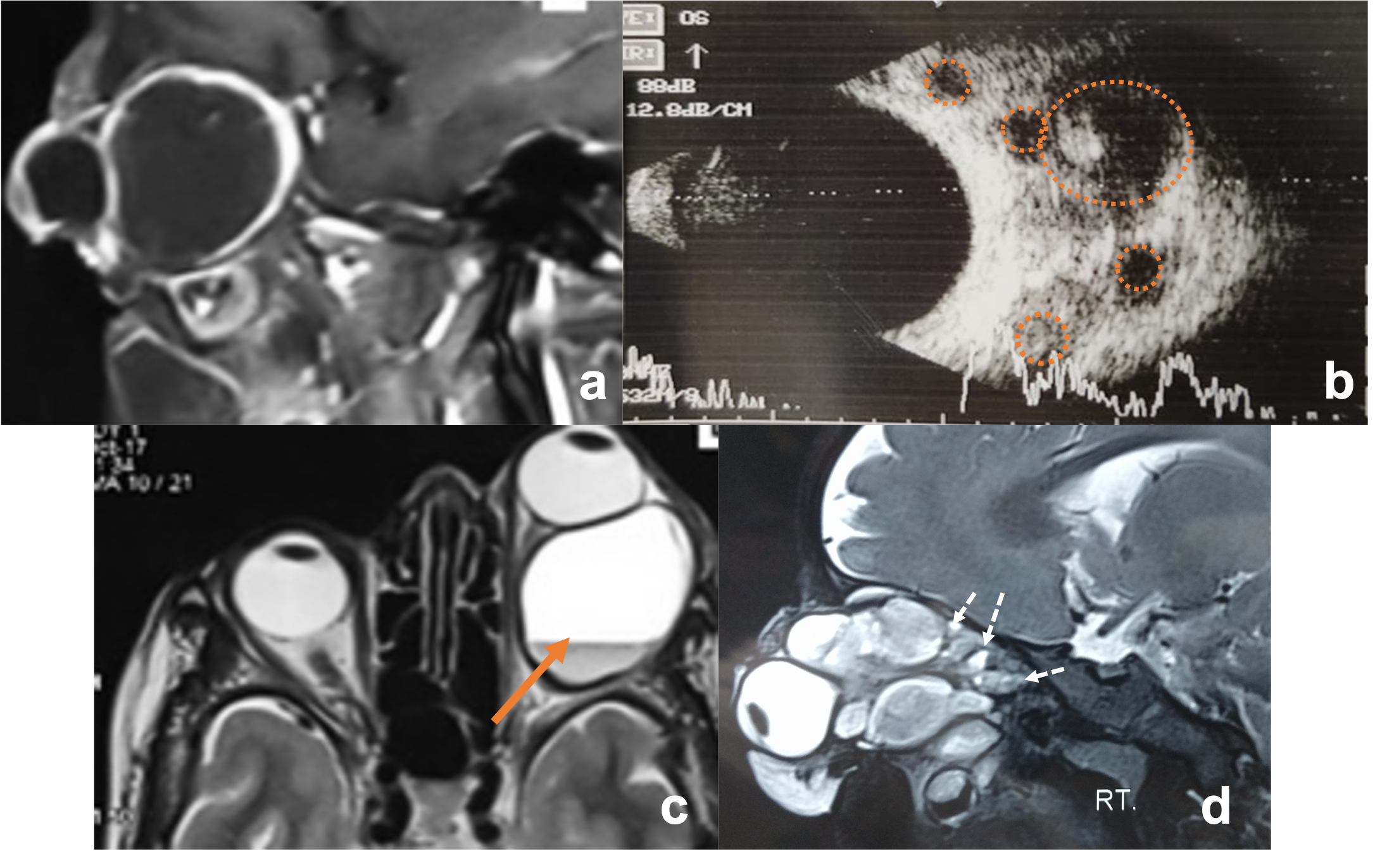
a,c Macrocystic lymphatic malformation. Sagittal T1 WI of left orbit showing large cyst with hypointense signal and Axial T2 WI MRI showing large cyst with fluid-fluid levels(haemorrhagic content hyperintense on T2 signal(orange arrow) with septa of hypointense fluid) b) USG orbit demostrates MIXED variant lymphatic malformation, with multiple cyst (orange circles) d) Saggital T2 WI MRI of orbit showing multiple cyst with hyperintesne signal (microcyst marked with white arrow)
File history
Click on a date/time to view the file as it appeared at that time.
| Date/Time | Thumbnail | Dimensions | User | Comment | |
|---|---|---|---|---|---|
| current | 01:13, June 1, 2021 | | 2,191 × 1,372 (2.45 MB) | SAHIL.AGRAWAL (talk | contribs) |
You cannot overwrite this file.
File usage
The following page uses this file: