File:Macular I CNV.jpg
From EyeWiki
Size of this preview: 800 × 534 pixels. Other resolution: 1,505 × 1,004 pixels.
Original file (1,505 × 1,004 pixels, file size: 88 KB, MIME type: image/jpeg)
Summary
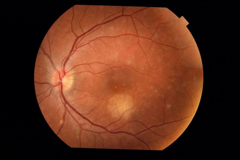
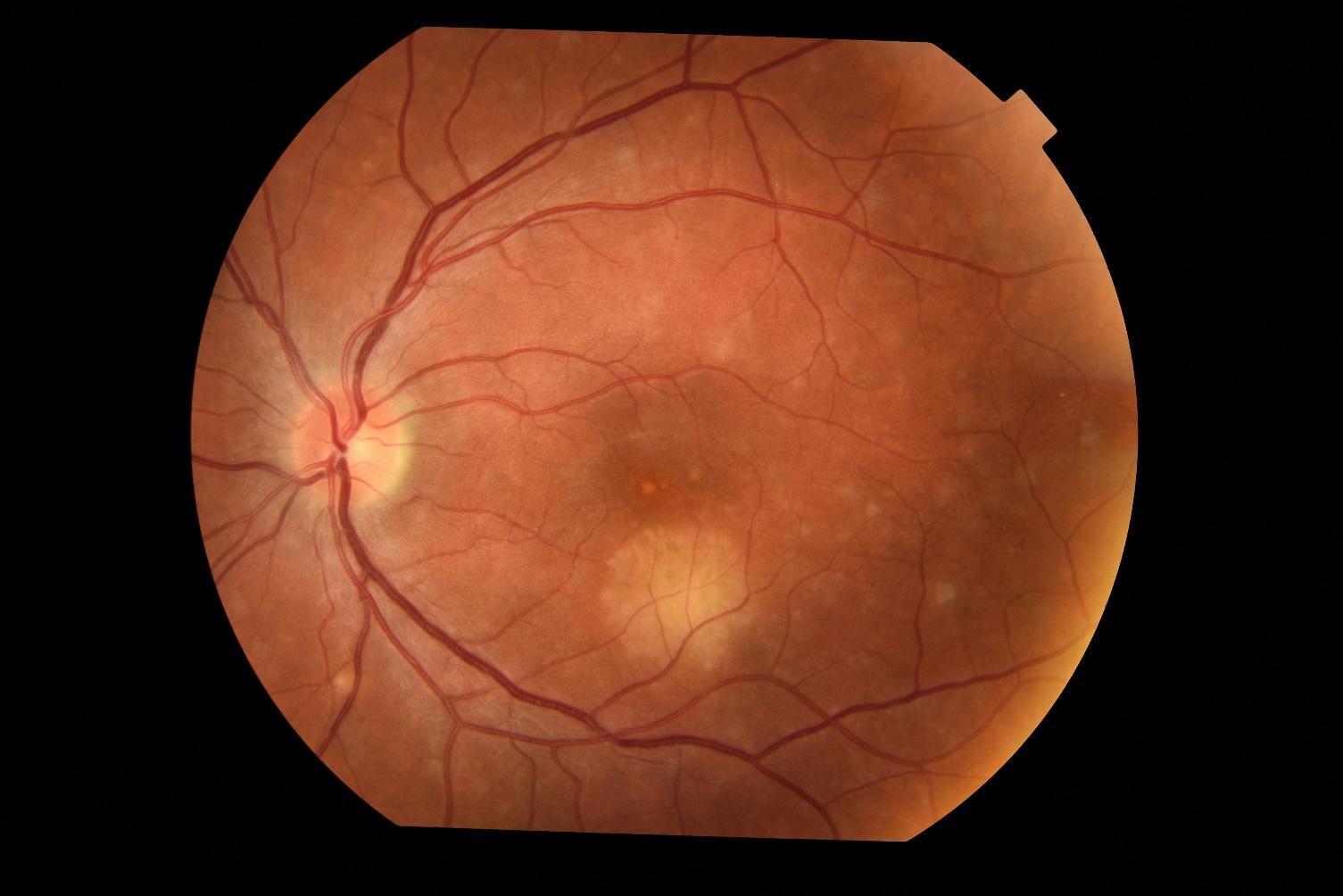
Figure 1: Color fundus photo of the left eye in a patient who described “spots” in the vision. The I-CNV appears as a creamy plaque with discrete borders inferior to the foveal center and under the vessels of the inferotemporal arcade. There are also numerous discrete hypopigmented lesions throughout the macula and posterior pole.
File history
Click on a date/time to view the file as it appeared at that time.
| Date/Time | Thumbnail | Dimensions | User | Comment | |
|---|---|---|---|---|---|
| current | 12:38, November 28, 2021 | | 1,505 × 1,004 (88 KB) | Justin.Yamanuha (talk | contribs) | Figure 1: Color fundus photo of the left eye in a patient who described “spots” in the vision. The I-CNV appears as a creamy plaque with discrete borders inferior to the foveal center and under the vessels of the inferotemporal arcade. There are also numerous discrete hypopigmented lesions throughout the macula and posterior pole. |
You cannot overwrite this file.
File usage
The following page uses this file: