File:Medulloepithelioma Combination Figure from IJO.jpg
Original file (845 × 1,039 pixels, file size: 145 KB, MIME type: image/jpeg)
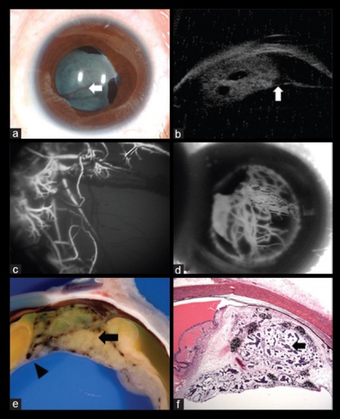
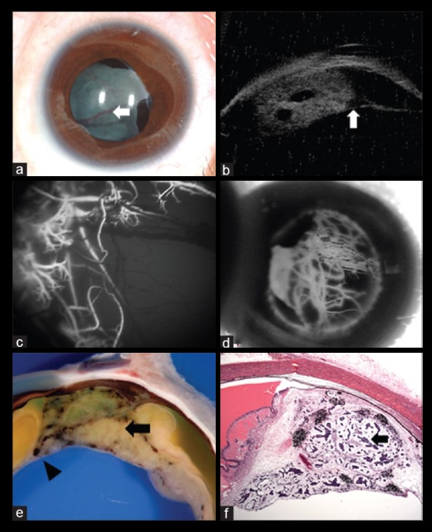
Figure 8 - A 5-year-old girl with (a) leukocoria was noted to have iris neovascularization and retrolental neoplastic cyclitic membrane with vessels (arrow) arising from the ciliary body. (b) Ultrasound biomicroscopy depicted the ciliary body mass with intratumoral cysts, lens subluxation and neoplastic cyclitic membrane (arrow). Fluorescein angiography in the (c) early and (d) late frames highlight the vessels in the retrolental membrane. Following enucleation, the (e) gross specimen reveals an amelanotic ciliary body tumor (arrow) with pigmented areas and with neoplastic cyclitic membrane (arrowhead). Histopathology (f) shows tubules and nests of basophilic neoplastic cells (black arrow) surrounded by loose mucopolysaccharide rich matrix. (Hematoxyline eosin stain; 2.5x magnification) – (Adapted with permission from Honavar et al. 2019 Indian Journal of Ophthalmology)
File history
Click on a date/time to view the file as it appeared at that time.
| Date/Time | Thumbnail | Dimensions | User | Comment | |
|---|---|---|---|---|---|
| current | 18:28, November 15, 2019 | | 845 × 1,039 (145 KB) | Mohammad.Sadiq (talk | contribs) | Figure 8 - A 5-year-old girl with (a) leukocoria was noted to have iris neovascularization and retrolental neoplastic cyclitic membrane with vessels (arrow) arising from the ciliary body. (b) Ultrasound biomicroscopy depicted the ciliary body mass with... |
You cannot overwrite this file.
File usage
The following page uses this file: