File:New Fig 1.png
From EyeWiki
Size of this preview: 790 × 599 pixels. Other resolution: 1,516 × 1,150 pixels.
Original file (1,516 × 1,150 pixels, file size: 3.02 MB, MIME type: image/png)
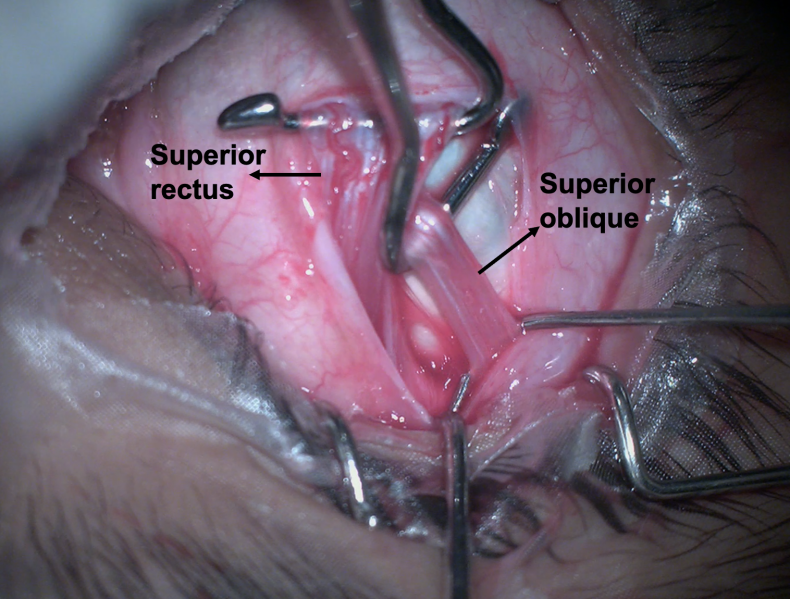
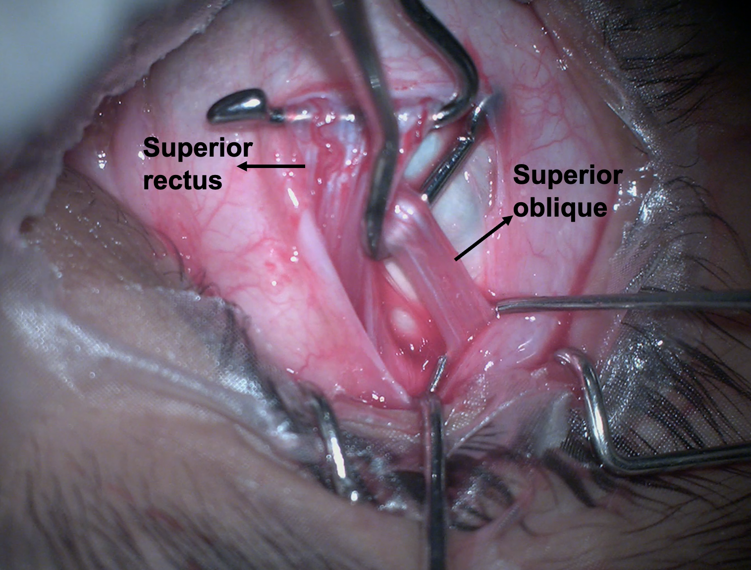
Figure 4- Intraoperative picture of the eye showing the superior rectus and superior oblique muscles. After reflection from the trochlea, the superior oblique passes beneath the superior rectus to get inserted on its lateral aspect.
File history
Click on a date/time to view the file as it appeared at that time.
| Date/Time | Thumbnail | Dimensions | User | Comment | |
|---|---|---|---|---|---|
| current | 00:25, May 31, 2023 | | 1,516 × 1,150 (3.02 MB) | Kirandeep.Kaur (talk | contribs) |
You cannot overwrite this file.
File usage
The following page uses this file: