File:Optic Nerve Glioma.png
From EyeWiki
Size of this preview: 800 × 373 pixels. Other resolution: 1,160 × 541 pixels.
Original file (1,160 × 541 pixels, file size: 659 KB, MIME type: image/png)
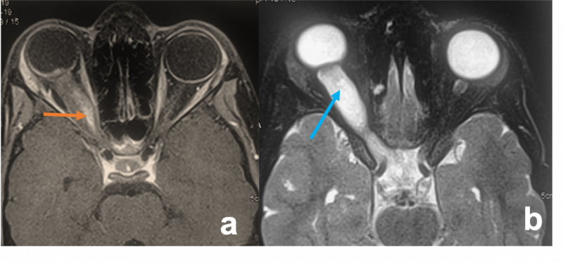
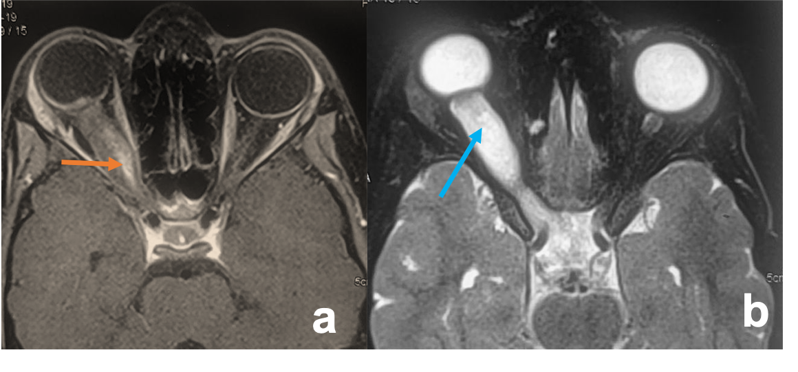
Optic Nerve Glioma a) Axial T1WI MR images demonstrate right optic nerve globular shaped mass of hypointense T1 and b) hyperintense T2 signal with mild post-contrast enhancement(orange arrow). The optic nerve cannot be made out separately. Note the presence of a small area of cystic degeneration inside the lesion (blue arrow).
File history
Click on a date/time to view the file as it appeared at that time.
| Date/Time | Thumbnail | Dimensions | User | Comment | |
|---|---|---|---|---|---|
| current | 04:36, June 1, 2021 | | 1,160 × 541 (659 KB) | SAHIL.AGRAWAL (talk | contribs) |
You cannot overwrite this file.
File usage
The following page uses this file: