File:Primary Angle Closure Using OCT in Asians.jpg
From EyeWiki
No higher resolution available.
Primary_Angle_Closure_Using_OCT_in_Asians.jpg (655 × 356 pixels, file size: 29 KB, MIME type: image/jpeg)
Summary
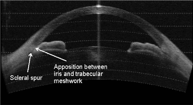
Anterior segment optical coherence tomography image of nasal and temporal angles showing apposition between the peripheral iris and angle wall anterior to the scleral spur Detection of Primary Angle Closure Using Anterior Segment Optical Coherence Tomography in Asian Eye. Ophthalmology 2007;114:33–39 © 2007
File history
Click on a date/time to view the file as it appeared at that time.
| Date/Time | Thumbnail | Dimensions | User | Comment | |
|---|---|---|---|---|---|
| current | 15:02, May 11, 2023 | | 655 × 356 (29 KB) | Tony.Ching.AAO (talk | contribs) | Anterior segment optical coherence tomography image of nasal and temporal angles showing apposition between the peripheral iris and angle wall anterior to the scleral spur Detection of Primary Angle Closure Using Anterior Segment Optical Coherence Tomography in Asian Eye. Ophthalmology 2007;114:33–39 © 2007 |
You cannot overwrite this file.
File usage
The following page uses this file: