File:Retinoblastoma.png
From EyeWiki
Size of this preview: 505 × 600 pixels. Other resolution: 847 × 1,006 pixels.
Original file (847 × 1,006 pixels, file size: 1.13 MB, MIME type: image/png)
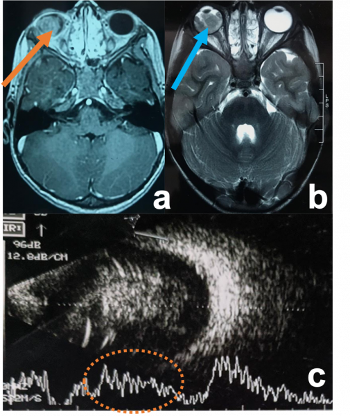
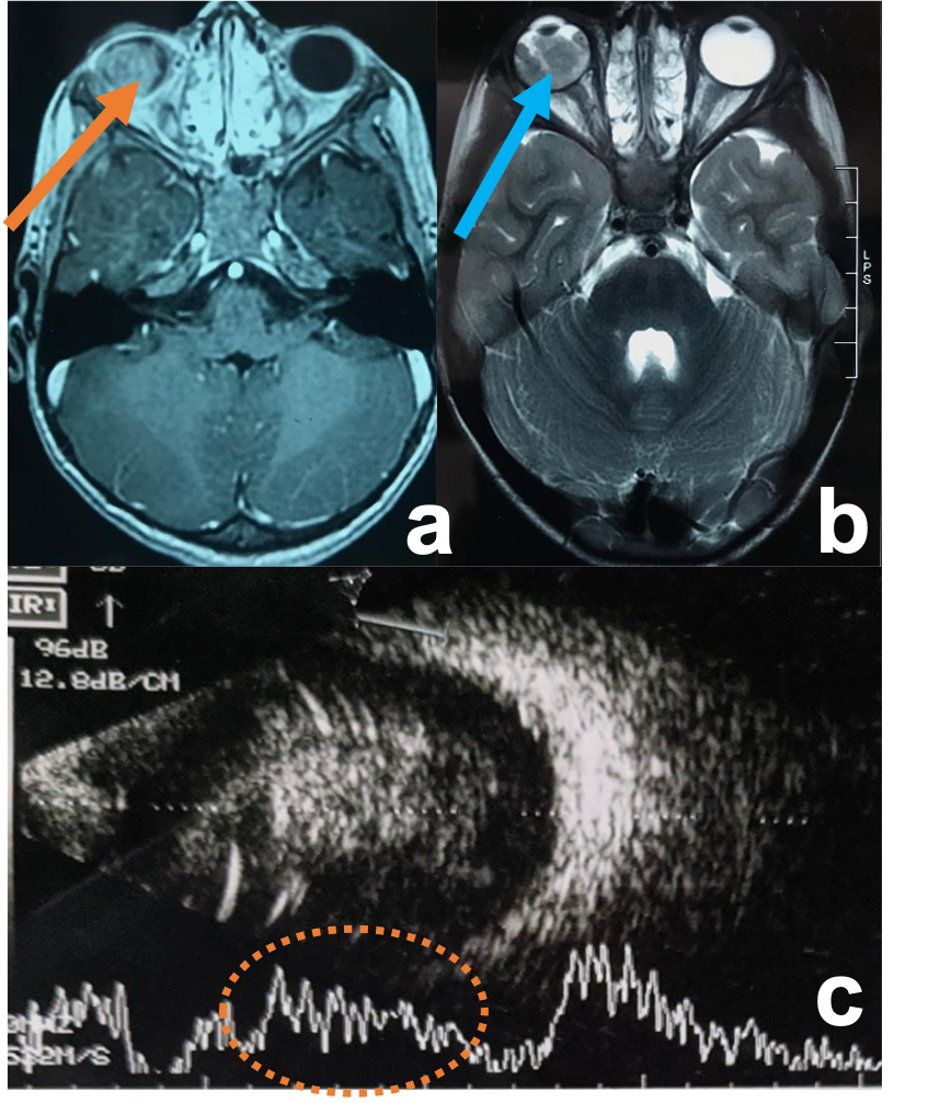
Retinoblastoma a) Contrast-enhanced T1WI axial MRI image showing an enhancing right intraocular mass with no extraocular extension (orange arrow) b) T2WI axial MRI image showing hypointense signal (blue arrow) c) USG (B/A) showing intraocular mass lesion with specks of calcification corresponding to the spikes of A-scan.
File history
Click on a date/time to view the file as it appeared at that time.
| Date/Time | Thumbnail | Dimensions | User | Comment | |
|---|---|---|---|---|---|
| current | 05:44, June 1, 2021 | | 847 × 1,006 (1.13 MB) | SAHIL.AGRAWAL (talk | contribs) |
You cannot overwrite this file.
File usage
The following page uses this file: