

File:Triangle-of-guillain-and-mollaret (1).jpg
Original file (1,000 × 1,024 pixels, file size: 194 KB, MIME type: image/jpeg)
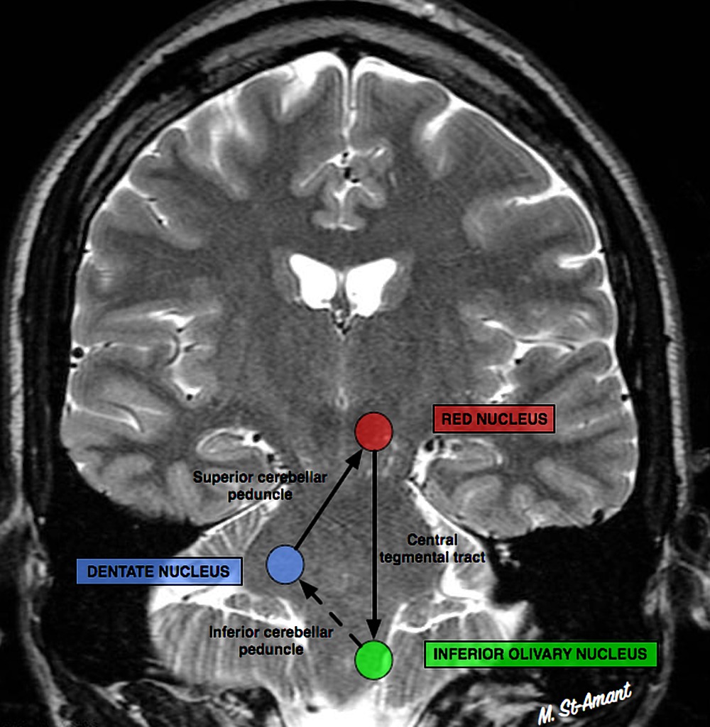
A schematic diagram of the dentate-rubro-olivary triangle (or Triangle of Guillain and Mollaret) over a coronal MRI of the brain. The dentate nucleus (blue circle) in the cerebellum sends signals along the superior cerebellar peduncle to the contralateral red nucleus (red circle) in the midbrain, which subsequently sends signals along the central tegmental tract to the inferior olivary nucleus (green circle) in the medulla. (Case courtesy of Dr Maxime St-Amant, https://radiopaedia.org/. From the case https://radiopaedia.org/cases/34774.)
File history
Click on a date/time to view the file as it appeared at that time.
| Date/Time | Thumbnail | Dimensions | User | Comment | |
|---|---|---|---|---|---|
| current | 12:57, October 9, 2018 | | 1,000 × 1,024 (194 KB) | Ore-ofe.Adesina (talk | contribs) | A schematic diagram of the dentate-rubro-olivary triangle (or Triangle of Guillain and Mollaret) over a coronal MRI of the brain. The dentate nucleus (blue circle) in the cerebellum sends signals along the superior cerebellar peduncle to the contralate... |
You cannot overwrite this file.
File usage
The following page uses this file:


.jpg)
.jpg&action=edit&redlink=1)
.jpg&action=edit)
.jpg&action=history)
.jpg&action=purge)
.jpg&oldid=37888)