File:Unilateral melanocytosis.jpg
Original file (2,083 × 1,086 pixels, file size: 359 KB, MIME type: image/jpeg)
Summary
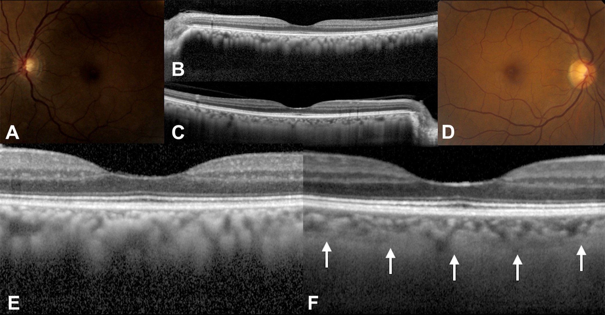
A 58-year-old African American woman with diffuse unilateral melanocytosis affecting the left eye (A) and normal clinical findings in the right eye (D). B, C, Horizontal optical coherence tomography (OCT) scans through the fovea show bilaterally a normal retina with no view of the sclerochoroidal interface in the study eye (B). Magnification of OCT scans shows a thickened choroid in the study eye (E) compared with the opposite eye (F), white arrows: sclerochoroidal junction). Choroidal Melanocytosis Evaluation with Enhanced Depth Imaging Optical Coherence Tomography, Ophthalmology, Volume 121 Issue 1 Pages 257-261 (January 2014)
File history
Click on a date/time to view the file as it appeared at that time.
| Date/Time | Thumbnail | Dimensions | User | Comment | |
|---|---|---|---|---|---|
| current | 16:03, May 17, 2023 | | 2,083 × 1,086 (359 KB) | Tony.Ching.AAO (talk | contribs) | A 58-year-old African American woman with diffuse unilateral melanocytosis affecting the left eye (A) and normal clinical findings in the right eye (D). B, C, Horizontal optical coherence tomography (OCT) scans through the fovea show bilaterally a normal retina with no view of the sclerochoroidal interface in the study eye (B). Magnification of OCT scans shows a thickened choroid in the study eye (E) compared with the opposite eye (F), white arrows: sclerochoroidal junction). Choroidal Melano... |
You cannot overwrite this file.
File usage
The following page uses this file: