File:Xanthelasma Cells.jpg
From EyeWiki
Size of this preview: 800 × 450 pixels. Other resolution: 1,366 × 768 pixels.
Original file (1,366 × 768 pixels, file size: 196 KB, MIME type: image/jpeg)
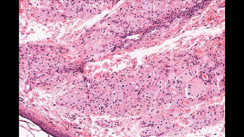
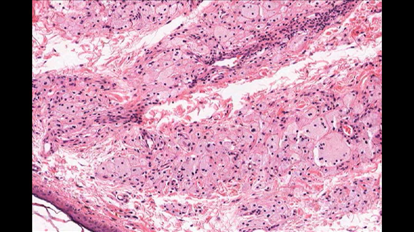
Image 2: Xanthelasma Cells, cutaneous manifestation of lipid laden foamy cells in the epidermis. They manifest as yellowish white plaque like lesions in the eyelid skin, commonly in the medial canthus. Image courtesy of Dr. William Morris, M.D.
File history
Click on a date/time to view the file as it appeared at that time.
| Date/Time | Thumbnail | Dimensions | User | Comment | |
|---|---|---|---|---|---|
| current | 17:15, September 20, 2018 | | 1,366 × 768 (196 KB) | Scdryden (talk | contribs) | Image 2: Xanthelasma Cells, cutaneous manifestation of lipid laden foamy cells in the epidermis. They manifest as yellowish white plaque like lesions in the eyelid skin, commonly in the medial canthus. Image courtesy of Dr. William Morris, M.D. |
You cannot overwrite this file.
File usage
The following page uses this file: