All content on Eyewiki is protected by copyright law and the Terms of Service. This content may not be reproduced, copied, or put into any artificial intelligence program, including large language and generative AI models, without permission from the Academy.
ICD 11: BD93.1Y: Lymphoedema secondary to other specified cause
Overview
Morbihan disease (MD), also known as solid persistent facial edema, lymphedema rosacea, morbus Morbihan and Morbihan syndrome, is a rare condition characterized by chronic, progressive, non-pitting edema (+/- erythema) of the upper two-thirds of the face, notably the periorbital tissue, forehead, glabella, nose, and cheeks, that may result in facial disfigurement and visual field narrowing [1][2][3]
History of the disease
MD was first observed in the 1950s. [4][5] It was named after Morbihan, a department in Brittany, France where the findings were described by a dermatologist, Dr Robert Degos.
Anatomy
MD affects the upper two thirds of the face, including:
Forehead
Glabella
Periorbital region: both preseptal and pretarsal tissue
Cheeks
Nose
Pathogenesis
The cause of MD remains unknown. Many authors propose that MD is caused by lymphatic dysregulation, chronic inflammation, or both. The hypotheses fall under several categories:
There is an imbalance between lymphatic production and drainage [6][7]
Mast cells obstruct dermal lymphatics or cause dermal fibrosis. [8][9][3]
Peri and intra-lymphatic granulomas obstruct lymphatic drainage [10][11]
Chronic inflammatory mediators, released due to underlying autoimmune dysregulation or infection, cause vascular wall damage and breakdown of connective tissue within the dermis leading to persistent exudation and resultant edema.[12][13][14][15][16]
Contact urticaria, in response to topical irritants, triggers local inflammation resulting in insufficient lymphatic drainage in individuals with pre-existing lymphatic drainage defects[7]
The relationship between Morbihan disease, rosacea and acne has been theorized due to histopathologic similarities. [14] The limitation of this hypothesis, however, is that many patients with MD do not have rosacea or acne.[1][2][17] The association between rosacea, acne and MD remains unclear.
The most common histopathologic findings reported include:
Perivascular and perifollicular lymphocytic and histiocytic infiltration
Presence of mast cells
Perifollicular fibrosis
Dilated lymphatic channels in the dermis
Dermal edema
Histopathology of upper eyelid tissue in a patient with Morbihan Disease using hematoxylin and eosin (H&E) stain. Findings are non-specific. (A) 10x. Dermal edema with dilated lymphatics. (B) 10x. Perifollicular fibrosis, dermal edema, and dilated vessels. (C) 20x. Dilated lymphatics and dermal edema. (D) 40x. Dermal edema and mast cells. Slides provided by David Plemel, MD.The right upper lid skin biopsy. Hematoxylin and eosin stain. 10x magnification. Microscopic diagnosis of dermal fibroplasia (red arrow), telangiectasia (blue arrow), edema, and mild (patchy) perifollicular chronic inflammation (green arrow). Credit: Rona Silkiss, MD, FACS
Other, less commonly described, findings include:
Perifollicular and peri-lymphatic epithelioid granulomas
Lymphatic histiocytic infiltration
Telangiectasia
Dilated blood vessels
Plasma cells
Giant cells
Neutrophils
Increased collagen spacing and thickness
Sebaceous gland hypertrophy/hyperplasia
Chronic folliculitis
Dermal fibroplasia
Epidemiology
The incidence and prevalence of MD are unknown.
Risk Factors
Male > Female
Despite previous literature indicating MD affects women more than men, which would be in keeping with rosacea, recent studies have found that there are more cases described in men than in women. [1][3]
Middle age
Most common ages 40-60, although cases reported between ages 14-88.[1][3]
Ethnicity: Caucasian/white
Most of the cases described in the literature were of Caucasian individuals, followed by Asian individuals (Japanese, Chinese, Korean). Few reports in other ethnicities.[1]
Other: exposure to sun and woodworking dust.[27][28]
History
Patients note an insidious onset of upper facial swelling. Pertinent points on history include:
Possible visual impairment from increased lacrimation or mass effect causing ptosis and visual field narrowing [19][28][2]
Edema that is generally not position dependent, although may be described as worse in the morning [19]
May be associated with hot sensation of face, facial flushing [29][3], or psychosocial distress due to cosmetic disfigurement[1]
Physical examination
Physical examination findings include:
Preoperative photographs of a patient with Morbihan syndrome (above). There is decreased periocular soft tissue swelling after upper eyelid blepharoplasty with injection of steroids into all four eyelids (below). Photographs provided by Karim G Punja, MD. Patient consent was obtained for the publication of these photos on EyeWiki.Non-pitting, solid edema affecting the upper two-thirds of the face.
Locations most commonly involved include eyelids, forehead, glabella, and cheeks[10][18]
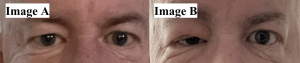
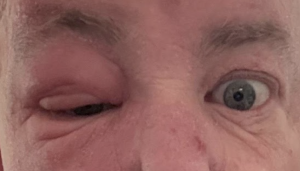
Image A was taken six months before image B, demonstrating progression of unilateral Morbihan’s disease. Treatment medications included dexamethasone 0.1%, methyl prednisone 4 mg dose pack, doxycycline hyclate 100 mg, ofloxacin 0.3%, amoxicillin-clavulanate 875–125 mg, prednisone 20 mg, tobramycin-dexamethasone 0.3%. Credit: Clinical photographs taken by Rona Z Silkiss, MD, FACS.Solid edema associated with Morbihan's disease commonly occurs on both sides of the face in a symmetrical pattern; rare variations include unilateral patterns documented three times in previous case presentations.[26]A unilateral presentation of Morbihan's disease is reported here. Active flare up experienced by the patient seven months after the initial symptom presentation of MD. Credit: Clinical photographs taken by Rona Z Silkiss, MD, FACS.
The edema has been described as hard, or woody, with a smooth surface[27]
Edema may initially be pitting, and become non-pitting over time[1]
Erythema of the overlying skin.[1] Erythema is typically ill-defined, present in discrete patches, or solitary plaques.
Findings may be symmetric or asymmetric, unilateral or bilateral [1][11]
Typically, preserved visual acuity and eye exam within normal limits [20][23]
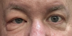
May lead to visual impairment / visual field narrowing due to ptosis from mass effect and lacrimation [19][28][2]Stable presentation of unilateral MD, eight months after initial flare-up. Credit: Clinical photographs taken by Rona Z Silkiss, MD, FACS.
Bilateral chemosis of the anterior segment has been reported in one case [1]
Signs of rosacea, telangiectasia, papules, pustules, granulomas, nodules [3][1][30]
Diagnosis
There are no diagnostic criteria for MD. It is a diagnosis of exclusion.[3] Investigations are used to rule out other causes of facial edema and are ordered at the discretion of the healthcare provider based on the patient’s specific presentation (see differential diagnoses below).
Laboratory test
The mainstays in investigations are bloodwork, radiographic imaging, and biopsy. Laboratory bloodwork can rule out systemic disease. Preoperative orbital computed tomography (CT) can be used to assess for orbital tumours. Biopsy of the skin can be used to rule out other dermatologic disease.
Investigations to consider
Rationale & Findings
Blood work
Note: Laboratory investigations are generally normal in MD
Complete blood count (CBC) for blood cell diseases
Inflammatory markers, C-reactive protein (CRP) or erythrocyte sedimentation rate (ESR), for inflammatory diseases
IgG4 for IgG4 disease
Angiotensin converting enzyme (ACE) and calcium for sarcoid
Albumin, bilirubin, creatinine, estimated glomerular filtration rate (eGFR) and alkaline phosphate (ALP) to assess for liver or renal anomalies.
Antinuclear antibody (ANA) and antineutrophil cytoplastic antibodies (ANCA) to screen assess for autoimmune markers
Thyroid stimulating hormone (TSH) and T3/T4 to assess for thyroid dysfunction.
Thyroid antibodies to assess for autoimmune thyroid disease.
Imaging
Orbital CT
To show preseptal, pretarsal and subcutaneous soft tissue swelling while ruling out orbital pathology.[20][1][23][31]
Chest x-ray or thoracic CT
To assess for hilar lymphadenopathy in sarcoidosis
Ultrasound and doppler flowmetry
Used in research studies to show insufficient lymphatic drainage[18]
Indocyanine green (ICG) lymphangiography
Used in research studies to identify locations of lymphatic vessels[7]
Melkersson-Rosenthal Syndrome (including all or part of the triad: facial palsy, facial edema, fissured tongue)
Congenital
Hypothyroidism, myxedema
SVC syndrome
Medications that can induced similar clinical signs: barbiturates, chlorpromazine, diltiazem, isotretinoin
Management
Systemic associations
Morbihan disease may be associated with rosacea, acne or both.[20][14][11][15]
Systemic manifestations
There are no systemic manifestations of MD.
Management of eyelid edema
There is no gold standard for the treatment of this rare disease.
Patients can be recommended avoidance potential triggers (see “prevention”).
Interventions result in variable clinical improvement. Recurrence or progression can be seen after treatment discontinuation.[1] A combination of interventions have been used with some success,[22][34][30][35][36] although a systematic review found no superior effect with combination therapy on outcomes and a greater risk of adverse effects.[1] Some case reports and case series have shown promising results.
Management in the literature
Most commonly used management strategies are highlighted in bold
Local
Intralesional steroid injection (triamcinolone) [15][22][24][3]Improvement of symptoms after steroid injection. Credit: Clinical photographs taken by Rona Z Silkiss, MD, FACS.
The authors would suggest injecting steroids periorbitally, deep to the orbicularis oculi muscle.
In a single case presentation, recurrent subcutaneous injection of a steroid was administered every six weeks until stabilization. After stabilization, repeat injections every 13 weeks led to cessation of edema.[26]
Lymphedema management including compression therapy, manual lymphatic drainage, skin care [7][34][37]
Other (less effective or less studied): Topical metronidazole and sulfur wash [1]
Median dose of 40mg PO daily for a duration of 6 months.[1]
Gradient dose of Isotretinoin; 30 mg, 60 mg, 80 mg. With consideration of sensitivity to the sun.[26]
Tetracyclines including doxycycline or minocycline[1][38][26]
Medial daily dose 200mg daily with duration correlating to treatment response; duration to partial response 3.0 month compared to 6.5 months for complete response [1]
The mechanism of action for omalizumab[26]. Adapted from Pelaia, Girolamo et al[40]. Omalizumab binds to free immunoglobulin E (IgE) so that it hinders binding to IgE receptors (FcεRI). Another alternative mechanism of omalizumab is that this medication can decrease effective IgE receptors shown on the mast cells and basophils, resulting in decreased swelling.
Janus activated kinase (JAK) inhibitors, such as Tofacitinib and Baricitinib[26]
The mechanism of action of JAK inhibitors[26]. Adapted from Shawky, Ahmed M, et al[41]. Janus kinase (JAK) inhibitors block the kinase domain of cytoplasmic non-receptor tyrosine kinases (JAKs), preventing the phosphorylation of STAT transcription factors. By disrupting the JAK-STAT pathway, they reduce cytokine-mediated signal transduction that drives inflammatory and autoimmune responses. This inhibition interferes with the pathogenic immune activation pathway, which promotes inflammation, immune response, and cancer.
Special considerations:
A review in 2019 found that oral steroids do not impact outcomes and are correlated with recurrences or progression [1]. Oral steroids such as prednisone acetate are often commonly prescribed[26].
Isotretinoin and tetracyclines should not be combined as this has been reported to increase the risk of idiopathic intracranial hypertension [42]
Authors have reported good outcomes using a combination of ultra-low-dose isotretinoin with antihistamines [35]
Paeoniflorin on nuclear factor kappa-light-chain-enhancer of activated B cells (NF-κB)[26] . Adapted from Sun[44], Liu et al[45], Yu et al[46]. Paeoniflorin inhibits the high mobility group box-1 (HMGB1), which plays a large role in activating the non-conical nuclear factor kappa-light-chain-enhancer of activated B cells (NF-κB) pathway. This pathway occurs as NF-κB-inducing kinase (NIK) activates NF-κB2 precursor protein, p100, leading to formation of p52/RelB dimer. The p52/RelB dimer is translocated to the cell nucleus leading to changes in gene expression and subsequent processes promoting inflammation and immune response.
Facial exercises to reduce periorbital and cheek swelling, such as movements of the tongue, facial muscles, neck and shoulder[26]
Successful treatments of Morbihan's disease patients from 2014 to 2021 by literature review published in "Successful treatment of a Morbihan's disease patient after a therapeutic challenge: A case report and comprehensive literature review" by Bazargan et al.[47]
Hattori, Y. et al.
2021
M
32
NA
Surgical Lymphaticovenous anastomosis
–
No recurrence after 1 year of follow-up
Donthi, D. et al.
2021
M
67
Mild spongiosis with periadnexal mixed infiltrate and dermal edema. The dermis showed perivascular and perifollicular lymphoplasmacytic inflammation, solar elastosis, and superficial dermal telangiectasia. No granulomata were observed
2 cases with a combination of 2.5% hydrocortisone cream, brimonidine 0.33% gel, metronidazole gel, and doxycycline
3 months
No recurrence after 1 year of follow-up
M
50
Dermis with perivascular and perifollicular lymphoplasmacytic inflammation, solar elastosis, and superficial dermal telangiectasias. No granulomata were observed
Cinar, G.N. et al.
2021
M
18
NA
Complete decongestive therapy for 24 sessions
8 weeks
NA
Yvon, C. et al.
2020
M: 8
F: 2
Mean age: 67
Features of inflammation and vascular dysfunction, are highly suggestive of a rosacea histological picture complicated by chronic lymphoedema
Ten cases: the most effective treatments included oral isotretinoin, intralesional triamcinolone, and debulking surgery
NA
NA
Welsch, K. et al.
2020
M: 2
F: 2
Mean age: 63
NA
Four cases with ultralow dose isotretinoin (10 mg daily) and antihistamines
Mean: 14 months
NA
Pflibsen, L.R. et al.
2020
M
55
Chronic inflammation, some granulomas, and histiocytes
Surgical resection
–
Contralateral involvement after 3 years
Kutlay, S. et al.
2019
F: 2
51, 45
NA
Two cases with complete decongestive therapy for 10–15 sessions
NA
NA
Olvera, V. et al.
2019
M: 3
51
Granulomatous blepharitis
Three cases with high-dose isotretinoin
1 year
No recurrence after 1 year of follow-up
62
Superficial perivascular dermatitis, dermal edema, and chronic lymphoplasmacytic perifolliculitis
29
Superficial and deep periadnexal dermatitis with lymphocytic infiltration
Kim, J.E. et al.
2019
NA
NA
Various extents of perivascular and perifollicular inflammation and dermal edema
Six cases with surgical eyelid reduction
–
NA
Kafi, P. et al.
2019
F
65
Non-specific inflammation consistent with rosacea
Omalizumab 450 mg
Monthly for 5 months
NA
Boparai, R.S. et al.
2019
F
40
Acanthotic epidermis with hypergranulosis and architectural disarray
Two cases with oral isotretinoin and prednisolone
NA
NA
M
61
Demodex and mild lymphocytic infiltrate in and around the follicles, as well as superficial vascular ectasia and prominent sebaceous glands
Cabral, F. et al.
2018
M
61
Edema between collagen fibers, dilated vessels with telangiectasias, and lymphocytic infiltrate
Oral isotretinoin and corticosteroid
NA
NA
Tsiogka, A. et al.
2018
M
44
Edema in upper dermis, discrete perivascular and periadnexal lymphocytic infiltrate without granulomatous reaction, and interstitial mast cells
Intralesional triamcinolone
Monthly for 4 months
No recurrence after 8 months of follow-up
Chaidemenos, G. et al.
2018
F
63
Perivascular and periadnexal lymphohistiocytic infiltrate, dermal edema, mild fibrosis, and sebaceous gland hyperplasia. Demodex mites within the follicular openings were also present
Doxycycline
8 months
NA
Yu, X. et al.
2017
M
42
Dermal telangiectasias and lymphocytic infiltration around the pilosebaceous glands
Marked edema and perivascular and perifollicular lymphocytic infiltration throughout the dermis
Minocycline 100 mg daily
4 months
NA
Fujimoto, N. et al.
2015
M
74
Marked dermal edema, perivascular and perisebaceous mononuclear cell infiltration, granulomatous reaction in the deep dermis, deep-reaching fibrosis, and numerous mast cells throughout the dermis
Minocycline
4 months
No recurrence after 8 months of follow-up
Literature review of novel therapeutics for Morbihan’s disease 2022–2024 meta-analysis published in "Unilateral presentation of Morbihan’s disease: a comprehensive case report and review" by Alvarez et al[26].
Author
Year
Sex
Age
Morbihan’s Disease Presentation
Histopathology
Treatment
Recurrence
Treatment Course
Article Link
Li, Z. et al.
2022
F
55
N/A
Hyperkeratosis of the epidermis, nodular inflammatory lesions in the dermis, epithelioid granuloma, and inflammatory infiltrating cells dominated by lymphocytes and histiocytes around skin appendages and blood vessels.
Prednisone acetate tablets 20 mg/d for 1 month.Tripterygium wilfordii polyglycoside 6 tablets/d (total 4 months of treatment)Total glucosides of paeony capsules administered 0.6 g 3 times per day (2 months after).
N/A
Duration of 5 monthsTransaminase levels significantly increased after prescribed prednisone acetate and triptyergium wilfordii polyglycoside.
Perivascular dermal edema with dilatation of lymphatic vessels and telangiectasia, mixed lymphocyte infiltrate (histiocytes, plasma cells, few eosinophils).
Infiltration of lymphocytes, histiocytes around the perifollicular area in the middle dermis. No neutrophil or giant cell infiltration. Proliferation of mast cells and mucin deposition around the hair follicles noted Alcian blue staining.
Dapsone.
Erythema and swelling of the right eyelid decreased.
It has been hypothesized that medical therapy often fails due to impaired local delivery systems at the site of chronic inflammation and interstitial edema in MD patients.[15] Erythema and inflammatory signs may respond to medications, but edema often persists.[15]Combining surgical debridement with anti-inflammatory medical therapy may improve treatment response.[15]
Future considerations for management include the use of immunosuppressant medications to target lymphocyte populations. Azathioprine and omazilumab have been suggested as potential therapies.[15][39][3]
Prevention
No modifiable risk factor has been identified.[3] Patients can be recommended avoidance of sun and irritating cosmetics as supportive to treatment.[16][18]
Prognosis
Without treatment, MD is unlikely to resolve spontaneously.[27] The condition is localized to the face and has no known systemic manifestations. MD is often refractory to treatment; however, most cases show at least partial response to conventional treatment.[1] A review article on the topic suggests patients may benefit from 4- to 6- months of tetracycline-based antibiotics with the risk of side effects weighed against the benefits of treatment.[1] Oral steroids were correlated with recurrence or progression. Male gender correlated with lack of complete response to treatment.[1] Patients who undergo debulking respond to treatment although response may be partial.[1] Approximately 10% of patients have recurrence or progression of disease.[1]
↑ 2.02.12.22.32.42.52.6Kim JE, Sim CY, Park AY, et al. Case Series of Morbihan Disease (Extreme Eyelid Oedema Associated with Rosacea): Diagnostic and Therapeutic Approaches. Ann Dermatol. 2019;31(2):196-200
↑Degos R, Civatte J, Beuve-Méry M. Nouveau cas d’œdème érythémateux facial chronique. Bull Soc Fr Dermatol Syph 1973;80:257
↑Schimpf A. Dermatitis frontalis granulomatosa. Derm Wschr 1956;133:120
↑ 6.06.1Smith LA, Cohen DE. Successful Long-term Use of Oral Isotretinoin for the Management of Morbihan Disease: A Case Series Report and Review of the Literature. Arch Dermatol. 2012;148(12):1395-1398
↑ 7.07.17.27.37.4Hattori Y, Hino H, Niu A. Surgical Lymphoedema Treatment of Morbihan Disease: A Case Report. Ann Plast Surg. 2021;86(5):547-550
↑ 8.08.1Veraldi S, Persico MC, Francia C. Morbihan syndrome. Indian Dermatol Online J 2013;4:122–4.
↑ 9.09.1Ramirez-Bellver JL, Perez-Gonzalez YC, Chen KR, et al. Clinicopathological and Immunohistochemical Study of 14 Cases of Morbihan Disease: An Insight Into Its Pathogenesis. Am J Dermatopathol. 2019;41(10):701-710.
↑ 10.010.110.2Nagasaka T, Koyama T, Matsumura K, Chen KR. Persistent lymphoedema in Morbihan disease: formation of perilymphatic epithelioid cell granulomas as a possible pathogenesis. Clin Exp Dermatol. 2008;33(6):764-7
↑Camacho-Martinez F, Winkelmann RK. Solid facial edema as a manifestation of acne. J Am Acad Dermatol 1990;22:129–30
↑Cribier B. Physiopathologie de la rosacée [Physiopathology of rosacea]. Ann Dermatol Venereol. 2014 Sep;141 Suppl 2:S158-64
↑ 14.014.114.214.3Kuhn-Régnier S, Mangana J, Kerl K, et al. A Report of Two Cases of Solid Facial Edema in Acne. Dermatol Ther (Heidelb). 2017;7(1):167-174
↑ 15.0015.0115.0215.0315.0415.0515.0615.0715.0815.0915.1015.11Carruth BP, Meyer DR, Wladis EJ, et al. Extreme Eyelid Lymphedema Associated With Rosacea (Morbihan Disease): Case Series, Literature Review, and Therapeutic Considerations. Ophthal Plast Reconstr Surg. 2017;33(3S Suppl 1):S34-S38
↑ 16.016.116.216.316.4Olvera-Cortés V, Pulido-Díaz N. Effective Treatment of Morbihan's Disease with Long-term Isotretinoin: A Report of Three Cases. J Clin Aesthet Dermatol. 2019;12(1):32-34
↑Chaidemenos G, Apalla Z, Sidiropoulos T. Morbihan disease: successful treatment with slow-releasing doxycycline monohydrate. J Eur Acad Dermatol Venereol. 2018;32(2):e68-e69.
↑ 18.018.118.218.318.4Wohlrab J, Lueftl M, Marsch WC. Persistent erythema and edema of the midthird and upper aspect of the face (morbus morbihan): evidence of hidden immunologic contact urticaria and impaired lymphatic drainage. J Am Acad Dermatol. 2005;52(4):595-602
↑ 20.020.120.220.320.420.520.6Bernardini FP, Kersten RC, Khouri LM, Moin M, Kulwin DR, Mutasim DF. Chronic eyelid lymphedema and acne rosacea. Report of two cases. Ophthalmology. 2000;107(12):2220-3
↑ 21.021.1Chalasani R, McNab A. Chronic lymphedema of the eyelid: case series. Orbit. 2010;29(4):222-6
↑ 22.022.122.2Kuraitis D, Coscarart A, Williams L, Wang A. Morbihan disease: a case report and differentiation from Melkersson-Rosenthal syndrome. Dermatol Online J. 2020;26(6)
↑ 23.023.123.223.323.423.5Lai TF, Leibovitch I, James C, Huilgol SC, Selva D. Rosacea lymphoedema of the eyelid. Acta Ophthalmol Scand. 2004;82(6):765-7
↑ 24.024.1Tsiogka A, Koller J. Efficacy of long-term intralesional triamcinolone in Morbihan's disease and its possible association with mast cell infiltration. Dermatol Ther. 2018;31(4):e12609
↑ 25.025.125.2Vasconcelos RC, Eid NT, Eid RT, Moriya FS, Braga BB, Michalany AO. Morbihan syndrome: a case report and literature review. An Bras Dermatol. 2016;91(5 suppl 1):157-159
↑ 27.027.127.227.327.4Hu SW, Robinson M, Meehan SA, Cohen DE. Morbihan disease. Dermatol Online J. 2012;18(12):27
↑ 28.028.128.228.3Ranu H, Lee J, Hee TH. Therapeutic hotline: Successful treatment of Morbihan's disease with oral prednisolone and doxycycline. Dermatol Ther. 2010;23(6):682-685.
↑ 29.029.1Cabral F, Lubbe LC, Nobrega MM, Obadia DL, Souto R, Gripp AC. Morbihan disease: a therapeutic challenge. An Bras Dermatol. 2017;92(6):847-850
↑ 30.030.130.2Aboutaam A, Hali F, Baline K, Regragui M, Marnissi F, Chiheb S. Morbihan disease: treatment difficulties and diagnosis: a case report. Pan Afr Med J. 2018;30:226
↑ 31.031.131.2Pflibsen LR, Howarth AL, Meza Rochin A, Decapite T, Casey WJ 3rd, Mansueto LA. A Navajo Patient with Morbihan's Disease: Insight into Oculoplastic Treatment of a Rare Disease. Plast Reconstr Surg Glob Open. 2020;8(9):e3090.
↑ 32.032.1Messikh R, Try C, Bennani B, Humbert P. Efficacité des diurétiques dans la prise en charge thérapeutique de la maladie de Morbihan: trois cas [Efficacy of diuretics in the treatment of Morbihan's disease: three cases]. Ann Dermatol Venereol. 2012;139(8-9):559-563
↑West, B. A., Hoesly, P. M., LeBoit, P. E., & Homer, N. A. (2022). Cutaneous angiosarcoma presenting as bilateral periorbital edema. Orbit, 42(6), 621–623. https://doi.org/10.1080/01676830.2022.2056901
↑ 34.034.134.234.3Heibel HD, Heibel MD, Cockerell CJ. Successful treatment of solid persistent facial edema with isotretinoin and compression therapy. JAAD Case Rep. 2020;6(8):755-757
↑ 35.035.135.2Welsch K, Schaller M. Combination of ultra-low-dose isotretinoin and antihistamines in treating Morbihan disease - a new long-term approach with excellent results and a minimum of side effects [published online ahead of print, 2020 Feb 5]. J Dermatolog Treat. 2020;1-4
↑ 36.036.1Rebellato PR, Rezende CM, Battaglin ER, Lima BZ, Fillus Neto J. Syndrome in question. An Bras Dermatol. 2015;90(6):909-11
↑Kutlay S, Ozdemir EC, Pala Z, Ozen S, Sanli H. Complete Decongestive Therapy Is an Option for the Treatment of Rosacea Lymphedema (Morbihan Disease): Two Cases. Phys Ther. 2019;99(4):406-410.
↑Okubo A, Takahashi K, Akasaka T, Amano H. Four cases of Morbihan disease successfully treated with doxycycline. J Dermatol. 2017;44(6):713-716
↑ 39.039.1Kafi P, Edén I, Swartling C. Morbihan syndrome successfully treated with omalizumab. Acta Derm Venereol 2019;99:677–678.
↑Pelaia G, Gallelli L, Renda T, et al. Update on optimal use of omalizumab in management of asthma. J Asthma Allergy. 2011;4:49–59. doi: 10.2147/JAA.S14520 .
↑Shawky AM, Almalki FA, Abdalla AN, Abdelazeem AH, Gouda AM. A comprehensive overview of globally approved jak inhibitors. Pharmaceutics. 2022;14(5):1001. doi: 10.3390/pharmaceutics14051001 .
↑Lee AG. Pseudotumor cerebri after treatment with tetracycline and isotretinoin for acne. Cutis. 1995 Mar;55(3):165-8
↑Yu X, Qu T, Jin H, Fang K. Morbihan disease treated with Tripterygium wilfordii successfully. J Dermatol. 2018;45(5):e122-e123.
↑Liu T, Zhang L, Joo D, Sun S-C. NF-κB signaling in inflammation. Sig Transduct Target Ther. 2017;2 (1):17023. doi: 10.1038/sigtrans.2017.23 .
↑Yu H, Lin L, Zhang Z, Zhang H, Hu H. Targeting NF-κB pathway for the therapy of diseases: mechanism and clinical study. Sig Transduct Target Ther. 2020;5(1):209. doi: 10.1038/s41392-020-00312-6 .
↑Bazargan AS, Dehnavi AZ, Dehghani A, Hesari KK,
Jafari P, Goodarzi A. Successful treatment of Morbihan’s disease patient after a therapeutic challenge: a case report and comprehensive literature review. J Gen Fam Med. 2024;25(4):232–236. doi: 10.1002/jgf2.690 .
The Academy uses cookies to analyze performance and provide relevant personalized content to users of our website.